Figures & data
Figure 1 Tumor cells and edema in the brains of rats with advanced F98 gliomas.
Notes: (A and B) Left hemisphere (two separate rats) and (C) right hemisphere. (A–C) mCherry red (tumor, red), anti-albumin (edema, green), and DAPI (nuclei, blue).
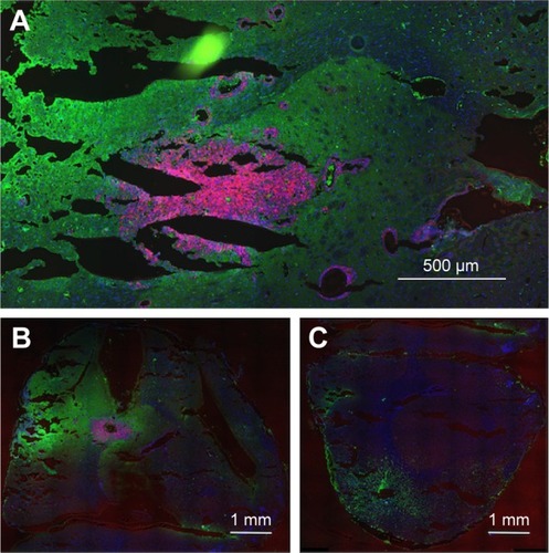
Figure 2 IV-injected AuNPs colocalize with F98 tumors. (A) Rat 1, left hemisphere, fluorescence; (B) Rat 1, left hemisphere, gold enhance; (C) Rat 1, right hemisphere, fluorescence; (D) Rat 1, right hemisphere, gold enhance; (E) Rat 2, left hemisphere, fluorescence; (F) Rat 2, left hemisphere, gold enhance; (G) Rat 2, right hemisphere, fluorescence; and (H) Rat 2, right hemisphere, gold enhance. (A–G) mCherry red (tumor, red), anti-albumin (edema, green), DAPI (nuclei, blue), and anti-CD31 (blood vessels, white) and (B–F) gold enhanced (gold stained, black).
Abbreviations: AuNPs, gold nanoparticles; CD31, cluster of differentiation 31; IV, intravenous.
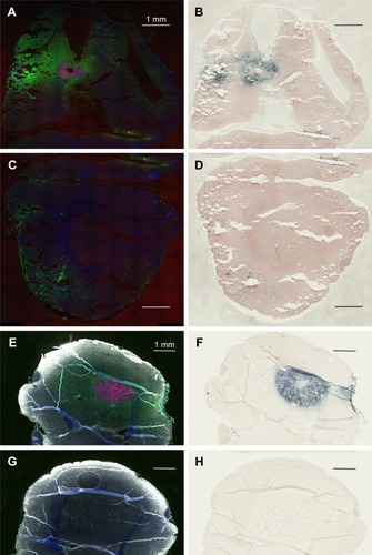
Figure 3 Complete lack of gold enhancement in saline-injected rats.
Notes: (A) Left hemisphere, fluorescence; (B) left hemisphere, gold enhance; (C) right hemisphere, fluorescence; and (D) right hemisphere, gold enhance. (A and C) mCherry red (tumor, red), anti-albumin (edema, green), DAPI (nuclei, blue), and anti-CD31 (blood vessels, white) and (B and D) gold enhanced (gold stained, black).
Abbreviation: CD31, cluster of differentiation 31.
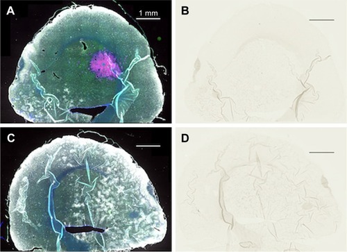
Figure 4 IV-injected AuNPs are associated with tumor cells lining blood vessels far from the main tumor mass.
Notes: Region far from the central tumor: (A) fluorescence and (B) gold enhanced. Saline control far from the central tumor: (C) fluorescence and (D) gold enhanced. (A and C) mCherry red (tumor, red), anti-albumin (edema, green), DAPI (nuclei, blue), and anti-CD31 (blood vessels, white) and (B and D) gold enhanced (gold stained, black).
Abbreviations: AuNPs, gold nanoparticles; CD31, cluster of differentiation 31; IV, intravenous.
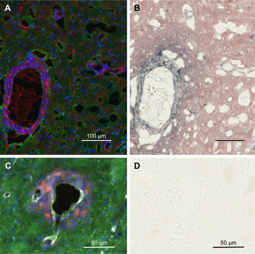
Figure 5 IV-injected AuNPs are found around tumor cells lining blood vessels and edema in peritumor region.
Notes: Peritumor region: (A) fluorescence, mCherry red (tumor, red), anti-albumin (edema, green), DAPI (nuclei, blue), and anti-CD31 (blood vessels, white) and (B) gold enhanced (gold stained, black). Scale bar, 50 μm.
Abbreviations: AuNPs, gold nanoparticles; CD31, cluster of differentiation 31; IV, intravenous.
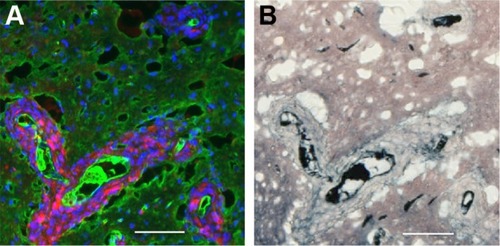
Figure 6 Directly injected AuNPs are found in the white matter track and peritumor region but not in the main tumor region.
Notes: (A) Left hemisphere, fluorescence; (B) left hemisphere containing a brain tumor, gold enhanced; (B, inset) left hemisphere with no brain tumor, gold enhanced; (C) right hemisphere, fluorescence; (D) right hemisphere, gold enhanced; (D, inset) right hemisphere with no brain tumor; and (E) another animal, left hemisphere, stained for neurons (anti-Tuj 1, red fluorescence). (A and C) mCherry red (tumor, red), anti-albumin (edema, green), DAPI (nuclei, blue), and anti-CD31 (blood vessels, white) and (B, D, and F) gold enhanced (gold stained, black).
Abbreviations: AuNPs, gold nanoparticles; CD31, cluster of differentiation 31.
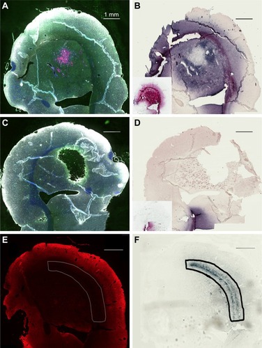
Figure 7 Directly injected AuNPs are largely found in the edema surrounding the tumor cells in peritumor region but not in and around the tumor cells themselves.
Notes: (A and C) Left hemisphere and (B and D) left hemisphere, gold enhanced. (A–C) mCherry red (tumor, red), anti-albumin (edema, green), DAPI (nuclei, blue), and anti-CD31 (blood vessels, white) and (B and D) gold enhanced (gold stained, black).
Abbreviations: AuNPs, gold nanoparticles; CD31, cluster of differentiation 31.
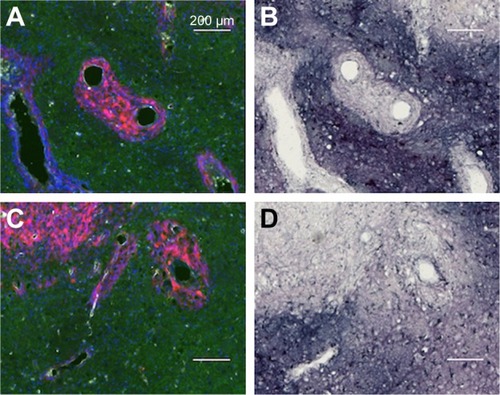
Figure 8 Quantification of the percentage of F98 containing BVs (F98 [+] BV) that also contained either a relatively large amount of AuNPs (AuNP++) or a relatively smaller amount of AuNPs (AuNP+) in the main tumor mass or in the peritumor region after either the direct infusion of AuNPs into the tumors or IV injection of AuNPs.
Abbreviations: AuNPs, gold nanoparticles; BV, blood vessel; IV, intravenous.
![Figure 8 Quantification of the percentage of F98 containing BVs (F98 [+] BV) that also contained either a relatively large amount of AuNPs (AuNP++) or a relatively smaller amount of AuNPs (AuNP+) in the main tumor mass or in the peritumor region after either the direct infusion of AuNPs into the tumors or IV injection of AuNPs.Abbreviations: AuNPs, gold nanoparticles; BV, blood vessel; IV, intravenous.](/cms/asset/fda45c64-81fe-4f95-8907-71e9e237e6cf/dijn_a_12193948_f0008_c.jpg)
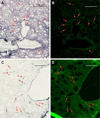
Figure 9 Uptake of AuNPs into brain microglia and astrocytes after injection of AuNPs directly into control brains (non-tumor).
Notes: (A and C) Gold enhanced (gold stained, black); (B) fluorescence, anti-Iba-1 (microglia, green); and (D) fluorescence, anti-GFAP (astrocytes, green).
Abbreviations: AuNPs, gold nanoparticles; GFAP, glial fibrillary acidic protein.