Figures & data
Figure 1 Physical characterization of Gal-PEI-SPIO nanoparticles.
Notes: (A) Schematic illustration of Gal-PEI-SPIO nano-encapsulated with siRNA and injected into mouse. (B, C) Diameters and zeta potentials of SPIO and Gal-PEI-SPIO nanoparticles. (D) Scanning electron microscope image of Gal-PEI-SPIO nanoparticles. Magnification ×200,000.
Abbreviations: SPIO, superparamagnetic iron oxide; Gal-PEI-SPIO, galactose-polyethylenimine-superparamagnetic iron oxide.
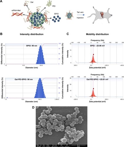
Figure 2 Agarose gel electrophoresis assay.
Notes: (A) siRNA bands with Gal-PEI-SPIOs at various siRNA:Fe (w/w) ratio. (B, C) Cell viability. Hepa1–6 cells were treated with Gal-PEI-SPIOs or Gal-PEI-SPIOs/siRNA at various ratios. (D) Heparin decomplexation assay. (E) siRNA stability assay.
Abbreviations: Gal-PEI-SPIO, galactose-polyethylenimine-superparamagnetic iron oxide; C, complexes; N, naked siRNA.
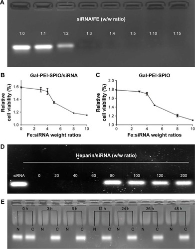
Figure 3 In vitro silencing efficacy of si-c-Met with Gal-PEI-SPIOs.
Notes: (A) The efficiency assessment of cellular uptake by flow cytometry in intro. Hepa1–6 cells were transfected with Gal-PEI-SPIO/Cy5-siRNA and equal Gal-PEI-SPIO/siRNA as control. (B) Quantitative PCR analyses the silencing efficacy of Gal-PEI-SPIO/si-c-Met by the relative c-Met mRNA levels in Hepa1–6 cells. (C, D) The Western blot assay shows the silencing efficacy of Gal-PEI-SPIO/si-c-Met in protein expression. *P<0.05.
Abbreviations: Gal-PEI-SPIO, galactose-polyethylenimine-superparamagnetic iron oxide; PCR, polymerase chain reaction.
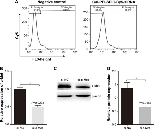
Figure 4 Cell proliferation inhibition assay.
Notes: (A, B) EdU stating. The proliferation inhibition of Gal-PEI-SPIO/siRNA was analyzed by EdU in Hepa1–6 cells. EdU-positive cells in the NC group were significantly more than in the c-Met group. ***P<0.0001.
Abbreviations: Gal-PEI-SPIO, galactose-polyethylenimine-superparamagnetic iron oxide; EdU, 5-ethylnyl-2′-deoxyuridine; NC, negative control.
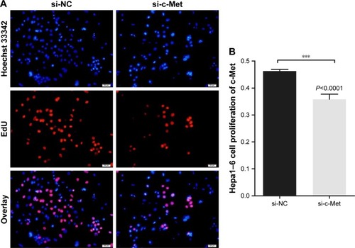
Figure 5 In vivo distribution of nanoparticles encapsulated with siRNA.
Notes: The Gal-PEI-SPIO/Cy5-siRNA complexions, Cy5-siRNA (0.5 mg siRNA/kg) were administered to the tumor-bearing mice via tail vein. At different time points (2 h, 8 h and 24 h), the fluorescence images were taken. (A) The distribution of Cy5-siRNA in tumor-bearing mice at different time periods. (B) The measurement of fluorescence intensity of Cy5-siRNA. In the Cy5-siRNA group, the fluorescence intensity was merely observed and significantly declined in the animal body at 24 h when compared with Gal-PEI-SPIO/Cy5-siRNA. *P<0.05.
Abbreviations: Gal-PEI-SPIO, galactose-polyethylenimine-superparamagnetic iron oxide; max, maximum; min, minimum.
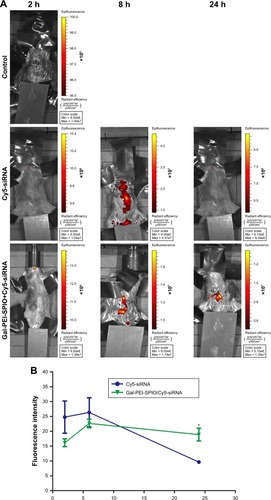
Figure 6 Tumor growth inhibition mediated by Gal-PEI-SPIO/si-RNA in vivo.
Notes: (A) The D-luciferin fluorescence intensity of mice bearing tumors. (B) The measurement of fluorescence intensity in different groups. (C) The tumor at final time point of treatment. (D) The ratio of liver:body weight of difference groups. (E) The volume of tumor. si-NC, Gal-PEI-SPIO/NC siRNA; si-c-Met; Gal-PEI-SPIO/si-c-Met siRNA; si-c-Met+M, Gal-PEI-SPIO/si-c-Met siRNA with addition of extra magnet. *P<0.05; **P<0.01.
Abbreviations: Gal-PEI-SPIO, galactose-polyethylenimine-superparamagnetic iron oxide; M, magnet; NC, negative control; NS, non-significance.
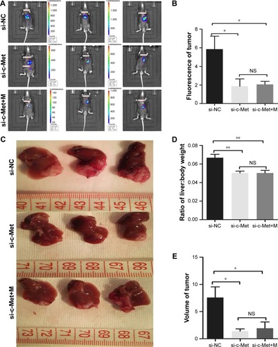
Figure 7 H&E staining and Prussian blue staining in tumor, renal and spleen tissue sections.
Notes: (A) Histological examination of the tumor, liver, spleen and kidney with H&E staining. (B) Prussian blue staining in tumor, renal and spleen tissue. Prussian blue spots of Gal-PEI-SPIO appearing as blue spots accumulated in tumor and spleen, but not in the kidney.
Abbreviations: H&E, hematoxylin-eosin; NC, negative control; M, magnet; Gal-PEI-SPIO, galactose-polyethylenimine-superparamagnetic iron oxide.
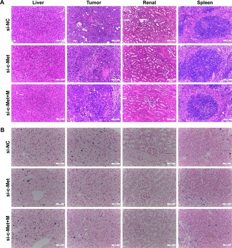
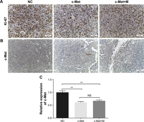
Figure 8 Immunohistochemistry assay.
Notes: (A) Histological examination of Ki-67. Ki-67-based qualification of cell proliferation in the tumor tissues (magnification ×400). (B) c-Met immunohistochemistry assay. IHC analysis of c-Met protein expression level in orthotopic tumors (magnification ×200). (C) The c-Met mRNA expression of tumor. Quantitative PCR of the tumor was used to assess the inhibition of tumor growth. **P<0.01.
Abbreviations: IHC, immunohistochemistry; M, magnet; NC, negative control; NS, non-significance; PCR, polymerase chain reaction.