Figures & data
Figure 1 Characterization of PDA and PDA–melittin nanoparticles.
Notes: (A) A scheme of the self-assembly process of PDA nanoparticles with bilayer structure. PCDA was self-assembled into colorless nanoparticles after probe sonication at 70°C. Polymerization of PCDA via a 1,4-addition reaction was achieved by UV irradiation at 254 nm, forming a blue polymer with alternating double-triple bond. (B) Transmission electron microscopy image of PDA nanoparticles. (C) UV spectrum of PDA nanoparticles. (D, E) The particle size distribution spectrum and the zeta potential distribution spectrum of PDA nanoparticles. (F, G) The particle size distribution spectrum and the zeta potential distribution spectrum of PDA–melittin nanoparticles. The test temperature was 25°C.
Abbreviations: PDA, polydiacetylene; PCDA, 10,12-pentacosadiynoic acid; NP, nanoparticle.
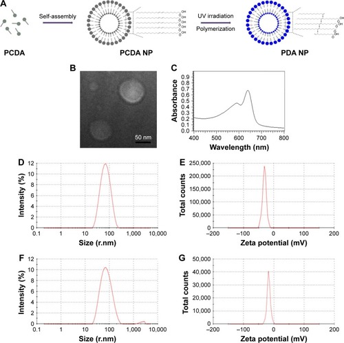
Figure 2 Interaction between PDA nanoparticles and melittin.
Notes: (A) Schematic presentation of the color variation after PDA binding with melittin. The solution color turns from blue to red after incubation at 37°C for 30 min. (B) The fluorescence alteration of PDA nanoparticles (20 mg/mL) after binding with melittin under a microscope at different time points. The fluorescence turned from none to red. (C) The red fluorescence intensity of PDA nanoparticles that were incubated with melittin of different concentrations at 37°C for 30 min. (D) The neutralization rate of PDA nanoparticles to melittin (5 μg/mL) at different ratios. (E) Centrifuged RBCs after incubated with melittin (5 μg/mL) combined with different concentrations of PDA nanoparticles. (F) Drug release of free melittin and PDA–melittin in vitro.
Abbreviations: PDA, polydiacetylene; RBCs, red blood cells.
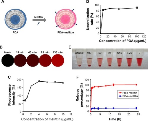
Figure 3 Characterization and immunization effect of PDA–melittin complex in vitro.
Notes: (A) Cellular uptake of PDA-melittin nanocomplex in DCs after incubation at 37°C with 5% CO2 atmosphere for 1 h. Scale bar: 5 μm. (B) Cell viability of 3T3 cells after treatment by PDA–melittin complex (PDA:melittin=10:1, the concentration of melittin is 6.25, 12.5, 25, 50, 100 μg/mL, respectively). (C) Cell viability of DCs after treatment by PDA–melittin complex (PDA:melittin=10:1, the concentration of melittin is 12.5 μg/mL). (D) The effect of PDA–melittin on the surface antigen expression of DCs.
Abbreviations: DCs, dendritic cells; PDA, polydiacetylene; MLT, melittin.
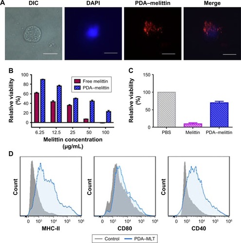
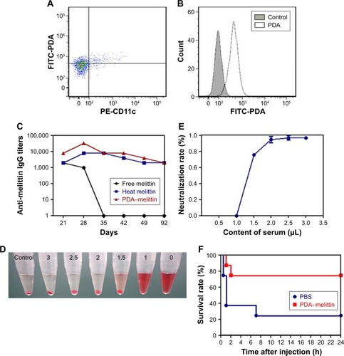
Figure 4 The immunization effect of PDA–melittin complex in vivo.
Notes: (A) The uptake of PDA nanoparticles to DCs in vivo by flow cytometry (scatter plot). (B) The uptake of PDA nanoparticles to DCs in vivo by flow cytometry (histogram). (C) Time course of anti-melittin IgG titers in free melittin treated mice (black circles), mice immunized with heat melittin (blue squares) or PDA–melittin complex (red triangles). (D) The hemolysis analysis of serum extracted from mice immunized with PDA–melittin complex. (E) The toxin neutralization effect of serum extracted from mice immunized with PDA–melittin complex. (F) Survival rate of mice challenged with melittin after immunized with PDA–melittin nanocomplex (red squares) and unimmunized (blue circles).
Abbreviations: PDA, polydiacetylene; FITC, fluorescein isothiocyanate; PE-CD11c, phycoerythrin-CD11C.