Figures & data
Figure 1 Effect of AuNPs on PDGF-induced VSMC migration.
Notes: The upper chamber, with VSMCs added in the presence of the indicated AuNPs or their corresponding vehicle ddH2O in (A) and 0.1 mg/mL citrate in (B), was assembled with the lower chamber filled with medium in the presence of vehicle, PDGF (10 ng/mL), and/or the indicated AuNPs. After incubation for 3 hours, VSMCs that had migrated to the underside of the filter membrane were photographed (left panels) and counted in HPF (magnification, 100×) under a phase-contrast light microscope (right panels). The results shown are representative of three to five independent experiments. **P<0.01, and ***P<0.001 versus PDGF+vehicle. Scale bar =100 μm.
Abbreviations: 3-EG AuNPs, 1-mercapto-(triethylene glycol)methyl ether functionalized AuNPs; 4-EG AuNPs, (1-mercaptoundec-11-yl)tetra(ethyleneglycol) functionalized AuNPs; AuNPs, gold nanoparticles; HPF, high-power field; pAuNPs, physically synthesized gold nanoparticles; PDGF, platelet-derived growth factor; std AuNPs, standard unconjugated AuNPs; VSMC, vascular smooth muscle cell; NS, nonsignificant; veh, vehicle.
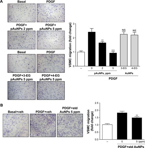
Figure 2 TEM and morphological analysis of VSMCs treated with pAuNPs.
Notes: VSMCs were incubated with pAuNPs for the indicated time intervals. (A) The subcellular distribution of the pAuNPs was analyzed by TEM. Scale bar =0.5 μm. (B) Cell morphology was analyzed by phase-contrast microscopy. Scale bar =100 μm.
Abbreviations: TEM, transmission electron microscopy; pAuNPs, physically synthesized gold nanoparticles; VSMC, vascular smooth muscle cell; ER, endoplasmic reticulum; M, mitochondria; N, nucleus.
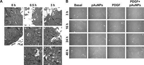
Figure 3 Effect of pAuNPs on viability, mitochondrial activity, and proliferation of VSMCs.
Notes: Cells were treated with PBS or PDGF (10 ng/mL) in the presence of vehicle or pAuNPs for the indicated time intervals. After incubation, cells were subjected to analysis by the LDH release (A), MTT (B), WST-1 (C), and BrdU incorporation (D) assays (n=3–5). *P<0.05, **P<0.01, and ***P<0.001 compared with each vehicle control. ##P<0.05 compared with basal control level. The arrows indicate a decrease in BrdU incorporation into DNA. Multiple groups were compared by one-way ANOVA.
Abbreviations: pAuNPs, physically synthesized gold nanoparticles; VSMC, vascular smooth muscle cell; PBS, phosphate-buffered saline; PDGF, platelet-derived growth factor; LDH, lactate dehydrogenase; ANOVA, analysis of variance.
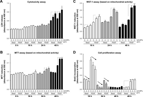
Figure 4 Effect of pAuNPs on PDGF signaling in VSMCs.
Notes: Cells were preincubated with Veh or pAuNPs (2 and 5 ppm) for 30 minutes, followed by PDGF (10 ng/mL) stimulation for (A) 10 minutes or (B) the indicated duration. After incubation, cells were collected, and protein phosphorylation and expression were determined by Western blotting (n=3). Similar data were quantitatively analyzed by densitometry. The phosphorylation level of each protein was calculated by the ratio of the expression level of phosphorylated protein to the expression level of the corresponding total protein. Data are expressed as fold changes relative to the basal level (untreated with PDGF). There is no significance between PDGF+vehicle control and each pAuNP treatment group.
Abbreviations: pAuNPs, physically synthesized gold nanoparticles; VSMC, vascular smooth muscle cell; PDGF, platelet-derived growth factor; Veh, vehicle.
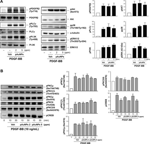
Figure 5 Gelatin zymography and Western blotting of MMP-2 expression and activity.
Notes: VSMCs were treated with Veh or pAuNPs (2 and 5 ppm) in the presence or absence of PDGF (10 ng/mL) at 37°C for 16 hours. The media were removed, centrifuged, and then analyzed by gelatin zymography and WB. The quantitation of MMP-2 activity based on similar zymographic results is shown in the lower panel (n=3).
Abbreviations: pAuNPs, physically synthesized gold nanoparticles; VSMC, vascular smooth muscle cell; PDGF, platelet-derived growth factor; MMP-2, matrix metalloproteinase-2; Veh, vehicle; WB, Western blotting.
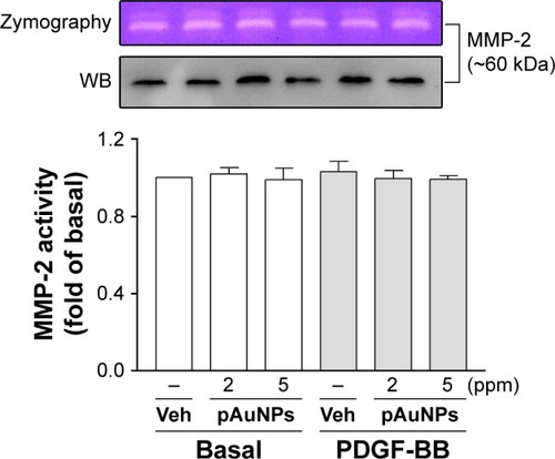
Figure 6 Effect of AuNPs on VSMC adhesion to collagen.
Notes: Cells were treated with vehicle or the indicated AuNPs for 30 minutes and allowed to adhere to collagen-precoated dishes in the presence or absence of PDGF (10 ng/mL) for 1 hour. VSMC adhesion was analyzed by (A–C) microscopy, MTT reduction, and (D) immunofluorescence microscopy and fluorometry (n=2–3). *P<0.05, **P<0.01, and ***P<0.001 compared with vehicle control. Scale bar =100 μm.
Abbreviations: 3-EG AuNPs, 1-mercapto-(triethylene glycol)methyl ether functionalized AuNPs; 4-EG AuNPs, (1-mercaptoundec-11-yl)tetra(ethyleneglycol) functionalized AuNPs; AuNPs, gold nanoparticles; pAuNP, physically gold nanoparticle; std AuNPs, standard unconjugated AuNPs; VSMC, vascular smooth muscle cell.
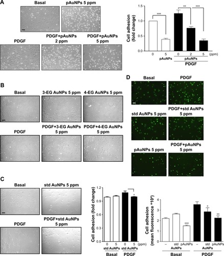
Figure 7 Effect of pAuNPs on collagen-induced protein tyrosine and FAK phosphorylation and actin cytoskeleton reorganization in VSMCs.
Notes: (A) Cells treated with vehicle or pAuNPs were allowed to adhere to collagen-precoated dishes for the indicated durations. Then, the cells were analyzed by Western blotting, and protein phosphorylation was quantified by densitometry (n=3–4). *P<0.05 compared with control. (B) Suspended VSMCs pretreated with vehicle (control) or pAuNPs (5 ppm) for 0.5 hours were allowed to adhere to collagen-precoated glass coverslips for an additional 1 hour. The cells were analyzed by immunofluorescence microscopy using anti-phosphotyrosine, p-FAK Ab, phalloidin-FITC, and DAPI as described in the Materials and methods section. Arrows indicate that stress fibers were formed in the control cells but disappeared in some pAuNP-treated cells. Scale bar =100 μm.
Abbreviations: pAuNPs, physically synthesized gold nanoparticles; VSMC, vascular smooth muscle cell; FAK, focal adhesion kinase; Ab, antibody; FITC, fluorescein isothiocyanate; DAPI, 4′,6-diamidino-2-phenylindole; Veh, vehicle.
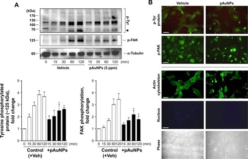
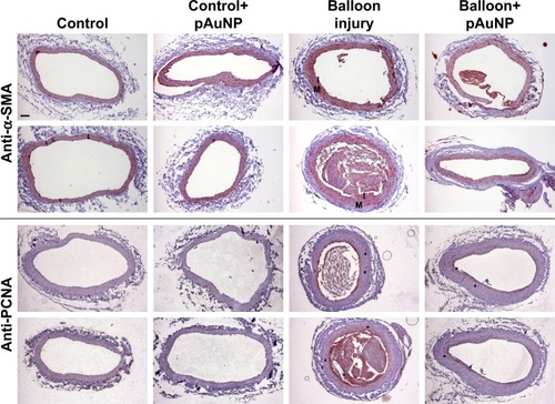
Figure 8 Effect of pAuNPs on the expression pattern of VSMCs in rat balloon-induced carotid artery.
Notes: Representative photomicrographs of α-SMA- and PCNA-stained sections of uninjured and balloon-injured carotid arteries of rats fed with or without the pAuNPs (five rats per group). The expression pattern of (proliferating) VSMCs in the sections was determined by immunohistochemistry using the Ab specific to α-SMA and PCNA. Red staining indicates positive regions. Arrows indicate proliferating VSMCs in the injured carotid arteries. Scale bar =100 μm. The images are representative of three independent experiments.
Abbreviations: pAuNPs, physically synthesized gold nanoparticles; PCNA, proliferating cell nuclear antigen; VSMC, vascular smooth muscle cell; α-SMA, α-smooth muscle actinin; M, media layer; I, intima; Ab, antibody.