Figures & data
Figure 1 Characterization of PLGA(Ag-Fe3O4).
Notes: (A) TEM image of Ag nanoparticles. (B) TEM image of Ag-Fe3O4 nanoparticles. (C) HRTEM image. (D) SEM image of PLGA(Ag-Fe3O4) covered on the planted tooth. (E) XRD. (F) FTIR spectrum. (G) Room temperature magnetic hysteresis loops of Ag-Fe3O4. (H) Ag-Fe3O4 multiple release behavior response to the magnetic field. It meant that Ag was stably bonded with Fe3O4.
Abbreviations: PLGA, poly (D, L-lactic-co-glycolic acid); SEI, secondary electron image; TEM, transmission electron microscopy; M, static magnetic field; emu, electromagnetic unit; H, magnetic field strength; Oe, oersted; SEM, scanning electron microscopy; XRD, X-ray powder diffraction; FTIR, Fourier transform infrared; HRTEM, high-resolution transmission electron microscopy; WD, working distance (the distance from the objective lens to focus point).
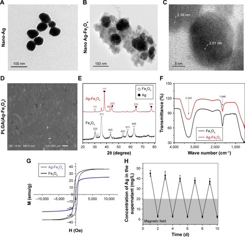
Figure 2 (A) Antibacterial results. PLGA(Ag-Fe3O4) nanoparticles were able to reduce Streptococcus mutans growth as nano-Ag, Ag-Fe3O4. **P<0.01. (B) Antibacterial activity test after washing. PLGA(Ag-Fe3O4) under magnetic field can be anchored on the tooth surface and exhibited a long-term antibacterial effect. (C) Results of S. mutans adhered to the samples surface. (D) SEM image of S. mutans adhered to the surface of different samples.
Abbreviations: Abs, absorbance; PLGA, poly (D, L-lactic-co-glycolic acid); SEM, scanning electron microscopy; M, static magnetic field; SEI, secondary electron image; WD, working distance (the distance from the objective lens to focus point).
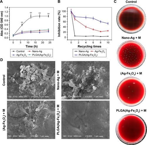
Figure 3 (A) Storage stability test result of PLGA(Ag-Fe3O4) nanoparticles. (B) ALP activity of osteoblasts in PBS, Fe3O4, or PLGA(Ag-Fe3O4) nanoparticles after 2, 4, and 6 days of differentiation culture; *P<0.05. **P<0.01. (C) Viability of osteoblasts incubated with different concentrations of samples for 24 hours. n=6; **P<0.01. (D) AgNOR staining in nucleoli of osteoblasts cultured with different nanoparticles. Original magnification 1,000×.
Abbreviations: M, static magnetic field; PLGA, poly (D, L-lactic-co-glycolic acid); ALP, alkaline phosphatase; AgNOR, silver staining for nucleolar organizer region.
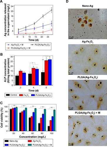
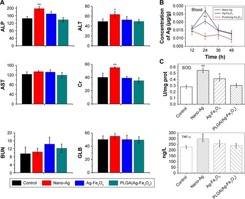
Figure 4 (A) Parameters of serum biochemistry profiles after administration of PLGA(Ag-Fe3O4) for 3 days. (B) The concentration of Ag in blood after treatment with nano-Ag and Ag-Fe3O4. Error bars represent standard error (n=5). (C) The degree of inflammation factors, including SOD and TNF-α in different samples-treated group. *P<0.05, **P<0.01.
Abbreviations: ALP, alkaline phosphatase; AST, aspartate aminotransferase; BUN, blood urea nitrogen; ALT, alanine aminotransferase; Cr, creatinine; GLB, globulin; PLGA, poly (D, L-lactic-co-glycolic acid); SOD, superoxide dismutase; TNF-α, tumor necrosis factor-α.
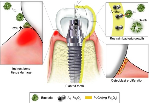
Scheme 1 Schematic diagram of PLGA(Ag-Fe3O4)-coated on dental implants.
Notes: Bacterial infection (such as the one by Streptococcus mutans) activates host immune response to produce ROS, leading to the destruction of the tooth supporting tissues (left). In the planted tooth (center) covered with PLGA(Ag-Fe3O4), bacterial adhesion ability weakened (top right). Thus, the immune system did not produce ROS and the microenvironment around the planted area stimulated osteoblast proliferation and improved the transplant success rate (bottom right).
Abbreviations: PLGA, poly (D, L-lactic-co-glycolic acid); ROS, reactive oxygen species; N, magnetic north pole; S, magnetic south pole.