Figures & data
Figure 1 Schematic process for preparation of micronized 10-hydroxycamptothecin-loaded folate-conjugated human serum albumin nanoparticles.
Abbreviations: nHPCT, micronized 10-hydroxycamptothecin; FA-HSA-nHCPT-NPs, micronized 10-hydroxycamptothecin-loaded folate-conjugated human serum albumin nanoparticles.
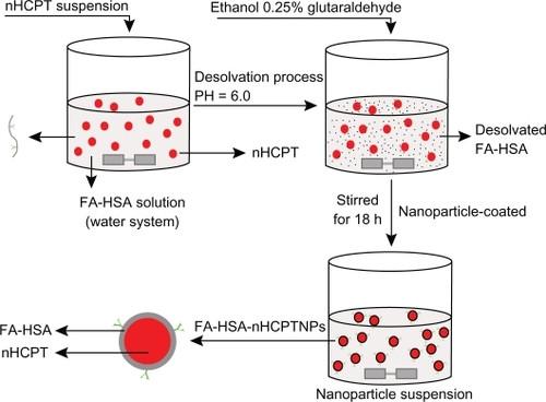
Figure 2 A) Determination of folate content conjugated with human serum albumin, a) N-hydroxysuccinimide ester of folate, b) tryptic hydrolysis of folate-conjugated human serum albumin nanoparticles, c) tryptic hydrolysis of human serum albumin nanoparticles; the particle size distribution of B) micronized 10-hydroxycamptothecin, C) micronized 10-hydroxycamptothecin-loaded folate-conjugated human serum albumin nanoparticles, D) HSA-nHCPT-NPs; the morphology observation of E) raw hydroxycamptothecin, F) micronized hydroxycamptothecin, G) micronized 10-hydroxycamptothecin-loaded folate-conjugated human serum albumin nanoparticles, and H) human serum albumin-loaded 10-hydroxycamptothecin nanoparticles, by scanning electron microscopy.
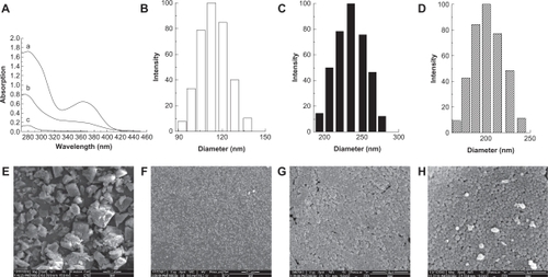
Figure 3 A) X-ray diffraction patterns, B) differential scanning calorimetry (DSC) curves, a) raw 10-hydroxycamptothecin, b) micronized 10-hydroxycamptothecin, and c) micronized 10-hydroxycamptothecin-loaded folate-conjugated human serum albumin nanoparticles.
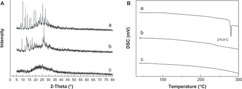
Figure 4 The release profiles of micronized 10-hydroxycamptothecin and micronized 10-hydroxycamptothecin-loaded folate-conjugated human serum albumin nanoparticles in vitro. (▴) Micronized 10-hydroxycamptothecin. (▵) Micronized 10-hydroxycamptothecin-loaded folate-conjugated human serum albumin nanoparticles.
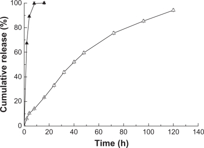
Figure 5 Confocal microscopy results. A549 cells (A and D) and SGC7901 cells (B, C, E, and F) were incubated with fluorescein isothiocyanate-labeled folate-conjugated human serum albumin nanoparticles or human serum albumin nanoparticles at indicated concentrations for four hours at 37°C. A) and B) 0.5 mg·mL−1 human serum albumin nanoparticles. C) 1 mg·mL−1 human serum albumin nanoparticles. D) and E) 0.5 mg·mL−1 folate-conjugated human serum albumin nanoparticles. F) 1 mg·mL−1 folateconjugated human serum albumin nanoparticles.
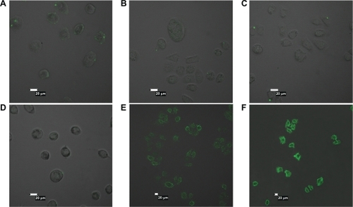
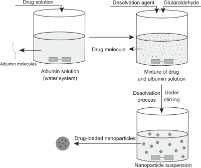
Figure 6 Schematic process of classical desolvation method.