Figures & data
Figure 1 Oleic acid–pluronic-stabilized iron oxide nanoparticles under transmission electronic microscopy.
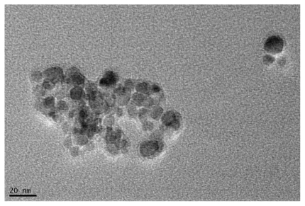
Figure 2 Fourier transfer infrared spectra by a Thermo Nicolet spectrometer. A) Oleic acid-coated iron oxide, B) pure Pluronic F-127, and C) oleic acid–pluronic stabilized iron oxide.
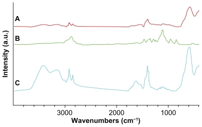
Figure 3 The crystallinity of pure Fe3O4 by X-ray diffractometry.
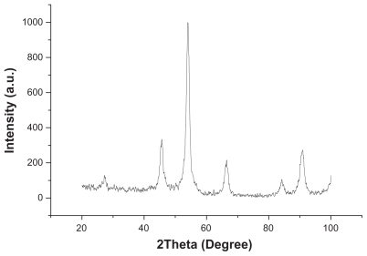
Figure 4 Hydrodynamic particle size distribution of daunorubicin-loaded magnetic nanoparticles in water measured by Zetasizer Nano ZS90 particle size analyzer.
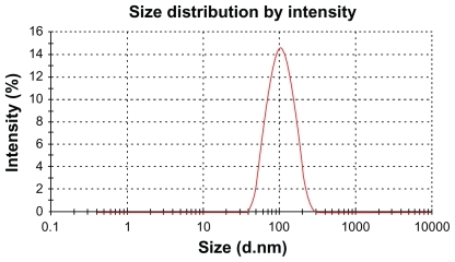
Table 1 Daunorubicin loading in magnetic nanoparticles (mean ± standard deviation, n = 3)
Figure 5 Release of daunorubicin from magnetic nanoparticles in vitro.
Note: Data are mean ± standard deviation (n = 3).
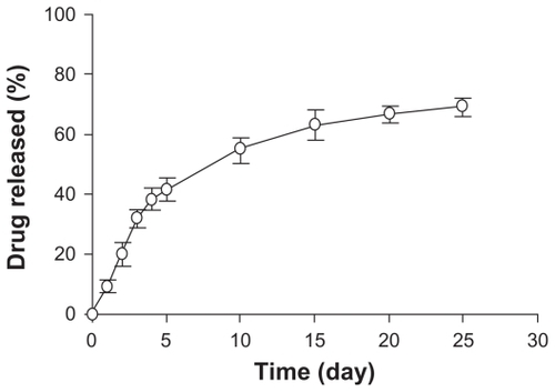
Figure 6 Magnetization as a function of field daunorubicin-loaded magnetic nanoparticles measured at 300 K.
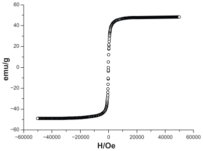
Figure 7 Cytotoxicity of K562 cells treated with different concentrations of DNRSol and DNR-MNPs by WST-1 assay.
Note: Data are mean ± standard deviation (n = 3).
Abbreviations: DNR-Sol, daunorubicin in solution; DNR-MNPs, daunorubicinloaded magnetic nanoparticles.
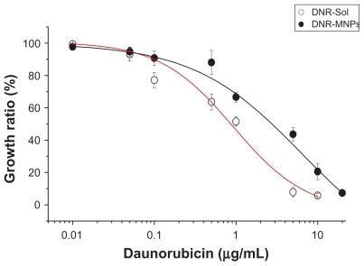
Figure 8 Cytotoxic effect of magnetic nanoparticles without drug on K562 cells.
Note: Data are mean ± standard deviation (n = 3).
Abbreviation: MNPs, magnetic nanoparticles.
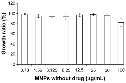
Figure 9 Cellular accumulation of DNR in K562 cells after treatment for 48 hours. A) control, B) DNR-Sol, and C) DNR-MNPs.
Abbreviations: DNR, daunorubicin; DNR-Sol, daunorubicin in solution; DNR-MNPs, daunorubicin-loaded magnetic nanoparticles.
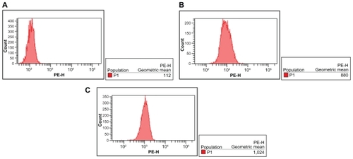
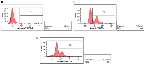
Figure 10 Effects of DNR-Sol and DNR-MNPs on apoptosis of K562 cells after treatment for 48 hours. A) control, B) DNR-Sol, and C) DNR-MNPs.
Abbreviations: DNR-Sol, daunorubicin in solution; DNR-MNPs, daunorubicin-loaded magnetic nanoparticles; MNPs, magnetic nanoparticles.