
Figures & data
Table 1 Biweekly body weight gain alterations induced in rats subjected to different injections of SDNPs in comparison with the rats received normal saline
Table 2 Changes on the average body weight (g) and the percentage of weight gain of rats subjected to different injections of SDNPs in comparison with rats received normal saline
Figure 1 Effect of SDNPs on body weight: twice-weekly changes in body weight gain with the number of SDNP injections.
Notes: *A significant difference (Student’s t-test, p<0.05) in comparison with the body weight gain of control rats. The decrease in the body weight gain of SDNP-treated rats in comparison with control rats is shown.
Abbreviations: SDNPs, silicon dioxide nanoparticles; w, week.
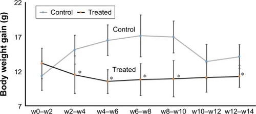
Table 3 Changes on the relative ratio of liver weight to body weight and the liver index of the control rats and those received 20, 35 or 50 injections of SDNPs (2 mg/kg bw)
Figure 2 Change in the liver index in correlation with the number of SDNP injections.
Note: The decrease in the liver index is directly proportional to the number of SDNP injections received by experimental rats.
Abbreviations: SDNPs, silicon dioxide nanoparticles; inj, injections.
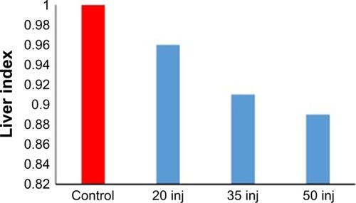
Table 4 Serum chemistry of control rats and those received 20, 35 or 50 injections of SDNPs (2 mg/kg bw)
Figure 3 Analyses of biochemical alterations induced by 20, 35 and 50 injections of 10-nm SDNPs (2 mg/kg body weight).
Note: *A significant difference (p-value, Student’s t-test) in comparison with the control group.
Abbreviations: ALP, alkaline phosphatase; ALT, alanine aminotransferase; AST, aspartate aminotransferase; HDL, high-density lipid; LDH, lactate dehydrogenase; SDNPs, silicon dioxide nanoparticles; inj, injections.
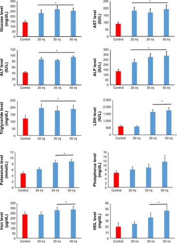
Table 5 Hematological analysis of control rats and those received 20, 35 or 50 injections of SDNPs (2 mg/kg bw)
Figure 4 Analyses of hematological alterations induced by 20, 35 and 50 injections of 10-nm SDNPs (2 mg/kg body weight).
Note: *A significant difference (p-value, Student’s t-test) in comparison with the control group.
Abbreviations: P-LCR, platelet larger cell ratio; SDNPs, silicon dioxide nanoparticles; WBCs, white blood cells; inj, injections.
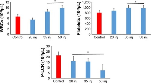
Figure 5 Microphotograph sections of the liver of control rats (A and B) and rats subjected to 50 injections of 10-nm SDNPs (C–H), stained by H&E.
Notes: (A) In control rats, the hepatic architecture demonstrates intact hepatic strands radiating outward from a central vein. (B) In control rats, hepatocytes display eosinophilic cytoplasm separated by vascular channels. The round nuclei dispersing smooth chromatin are shown. (C) Hydropic degeneration demonstrating swelling and cytoplasmic vacuolization. (D) Karyopyknosis showing nuclei shrinkage and chromatin condensation. (E) Infiltration of inflammatory cells demonstrating aggregation of inflammatory cells in the hepatic portal space. (F) Infiltration of lobular inflammatory cells (mainly lymphocytes) in the lobular hepatic strands. (G) Hyperplasia of Kupffer cells showing enlargement and hypertrophy of these defense cells. (H) Sinusoidal dilatation exhibiting widening of the capillaries lining the hepatic strands.
Abbreviations: H&E, with hematoxylin and eosin; SDNPs, silicon dioxide nanoparticles.
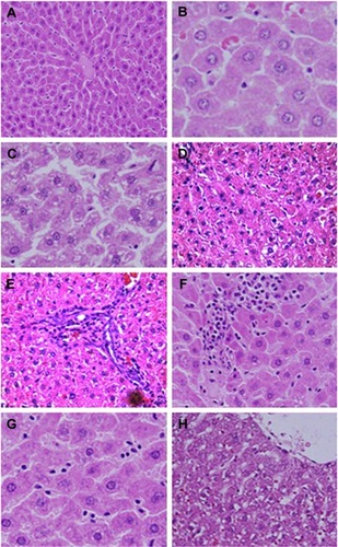
Table 6 Gene expression of drug-metabolizing enzymes in the hepatic tissues induced by SDNP exposure