Figures & data
Figure 1 TEM image of Mn0.5Zn0.5Fe2O4 nanoparticles.
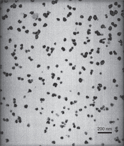
Figure 2 PEI-As2O3/MZF as seen by TEM (A) and SEM (B).
Abbreviations: PEI, polyethyleneimine;TEM, transmission electron microscopy; SEM, scanning electron microscopy.
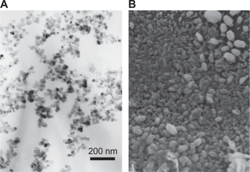
Figure 3 FTIR of PEI-As2O3/MZF: the top curve is As2O3/MZF and the bottom is PEI-As2O3/MZF.
Abbreviations: FTIR, Fourier transform infrared spectroscopy; PEI, polyethyleneimine.
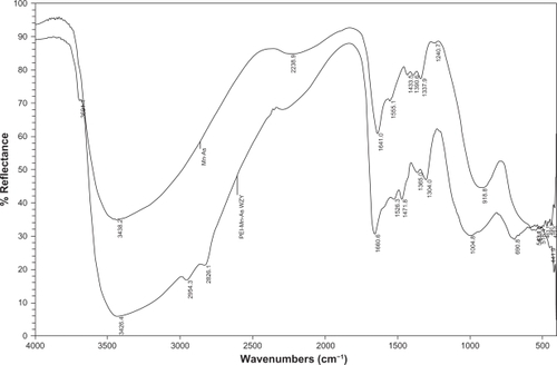
Figure 4 EDS results of PEI-As2O3/MZF.
Abbreviations: EDS, energy dispersive spectrometry.
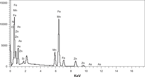
Figure 5 HepG2 cells observed under fluorescence microscopy (×400) 24 hours after transfection (A) and 48 hours after transfection (B).
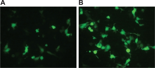
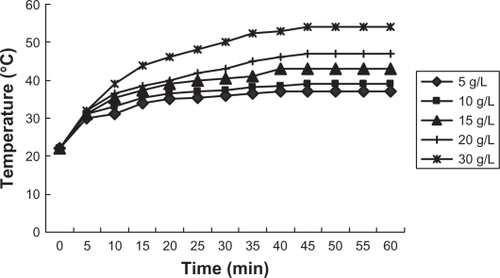
Figure 6 Heating test result of magnetic nanoliposomes.