Figures & data
Figure 1 Flow cytometric analysis of NIH/3T3 cells by double labeling with Annexin V-fluorescein isothiocyanate and propidium iodide. Unfixed cells from control and treated groups were labeled with Annexin V-fluorescein isothiocyanate and propidium iodide and then fixed and analyzed on a flow cytometer. Dual parameter dot-plot of propidium iodide-phycoerytrin (x-axis), Annexin V-fluorescein isothiocyanate fluorescence (y-axis) showing logarithmic intensity. Quadrants are viable cells Q3 (Annexin V-negative/propidium iodide-negative), early apoptotic cells Q4 (Annexin V-positive/propidium iodide-negative), and late apoptotic and necrotic cells Q2 (Annexin V-positive/propidium iodide-positive). Percentage of apoptotic cells are A) control cells, 1.5%; B) dimethyl sulfoxide control cells, 1.6%; C) Ma-Fol-modified magnetic nanoparticles, 2.5 μg/mL, 24 hours, 2.7%; D) Ma-Fol-modified magnetic nanoparticles 2.5 μg/mL, 48 hours, 2.8%; F) Ma-Fol-modified magnetic nanoparticles 4.5 μg/mL, 24 hours, 4.3%; G) Ma-Fol-modified magnetite nanoparticles 4.5 μg/mL, 48 hours, 4.7%; H) Ma-Fol-modified magnetic nanoparticles 9 μg/mL, 24 hours, 2.9%; I) Ma-Fol-modified magnetic nanoparticles 9 μg/mL, 48 hours, 3.1%.
Abbreviation: Ma-Fol, methacrylamido-folic acid.
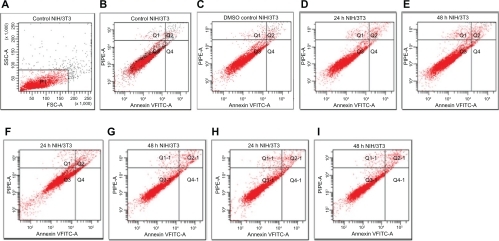
Figure 2 Flow cytometric analysis of 5RP7 (H-ras-transformed fibroblasts) by double labeling with Annexin V-fluorescein isothiocyanate and propidium iodide. Control cells were labeled with Annexin V-fluorescein isothiocyanate and propidium iodide, and then fixed and analyzed on a flow cytometer. Quadrants are viable cells Q3 (Annexin V-negative/propidium iodide-negative), early apoptotic cells Q4 (Annexin V-positive/propidium iodide-negative) and late apoptotic and necrotic cells Q2 (Annexin V-positive/propidium iodide-positive). Percentage of apoptotic cells are A) control cells, 1.1%; B) dimethyl sulfoxide control cells, 1.2%; C) Ma-Fol-modified magnetic nanoparticles 2.5 μg/mL, 24 hours 7.2%; D) Ma-Fol modified magnetic nanoparticles 2.5 μg/mL, 48 hours, 8.1%; E) Ma-Fol-modified magnetic nanoparticles 4.5 μg/mL, 24 hours, 10.5%; F) Ma-Fol-modified magnetic nanoparticles 4.5 μg/mL, 48 hours, 12.9 %.
Abbreviation: Ma-Fol, methacrylamido-folic acid.
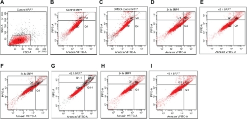
Figure 3 Transmission electron micrographs of Fe3O4 magnetic nanoparticles. A) 160,000×; B) 105,000×.
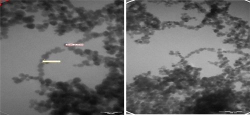
Figure 4 Transmission electron micrographs of NIH/3T3 cell (16,500×) not treated with folic acid-modified magnetic nanoparticles.
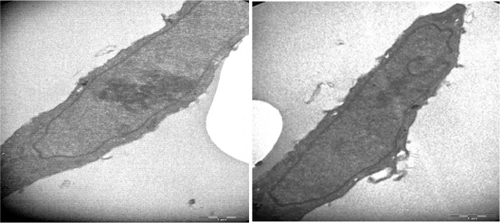
Figure 5 Transmission electron micrographs of a IH/3T3 cell. A) (6000×) treated with 4.5 μg/mL Ma-Fol-modified magnetic nanoparticles for 24 hours. B) (16,500×) treated with 4.5 μg/mL folic acid-modified magnetic nanoparticles for 48 hours.
Abbreviation: Ma-Fol, methacrylamido-folic acid.
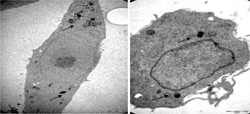
Figure 6 Transmission electron micrographs of 5RP7 cells not treated with folic acid-modified magnetic nanoparticles. C) 4200×; D) 8200×.
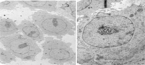
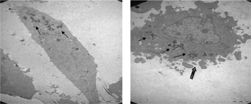
Figure 7 Transmission electron micrographs of 5RP7 cells; E) (8200×) (treated with 4.5 μg/mL folic acid-modified magnetic nanoparticles for 24 hours. F) (11,500×) treated with 4.5 μg/mL folic acid-modified magnetic nanoparticles for 48 hours leading to apoptotic blebbing.