
Figures & data
Table 1 Characteristics of SLNs (mean ± SD, n=3)
Figure 1 Photographs of tilmicosin-SLN and labeled tilmicosin-SLN (magnification was 10×40).
Notes: Left, optical microscopy image of tilmicosin-SLN; middle, optical microscopy image of labeled tilmicosin-SLN; and right, fluorescence microscopy image of labeled tilmicosin-SLN. The fluorescence microscopy image of labeled tilmicosin-SLN was obtained at the same area of normal light.
Abbreviation: SLN, solid lipid nanoparticle.
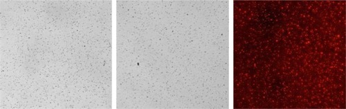
Table 2 MIC and MBC of native tilmicosin and tilmicosin-SLN
Figure 2 Sustained antibacterial activity of native tilmicosin and tilmicosin-SLN (drug concentration: 0.9 μg/mL).
Abbreviations: CFU, colony forming unit; SLN, solid lipid nanoparticle.
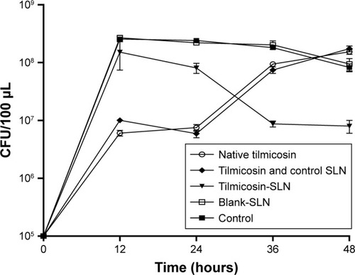
Table 3 Pharmacokinetic after s.c. injection in mice (mean ± SD, n=5)
Figure 3 Concentrations of tilmicosin in mammary glands after s.c. injection (60 mg/kg).
Abbreviations: s.c., subcutaneous; SLN, solid lipid nanoparticle.
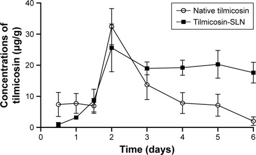
Figure 4 Fluorescence microscopy images of mammary gland slices.
Notes: Left, treated with labeled tilmicosin-SLN; middle, injection site; and right, treated with rhodamine B solution.
Abbreviation: SLN, solid lipid nanoparticle.
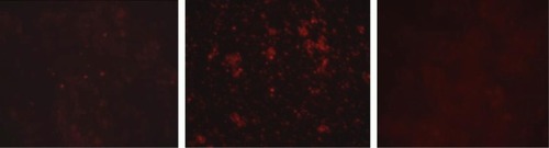
Table 4 The number of Streptococcus agalactiae in mammary glands (CFU/g) after they are infected with different inoculation quantities at 1 day (mean ± SD, n=4)
Figure 5 The number of Streptococcus agalactiae in mammary glands that were infected with 1×10⁷ CFU/100 µL per mammary gland at 1, 3, and 6 days (mean ± SD, n=5).
Abbreviation: CFU, colony forming unit.
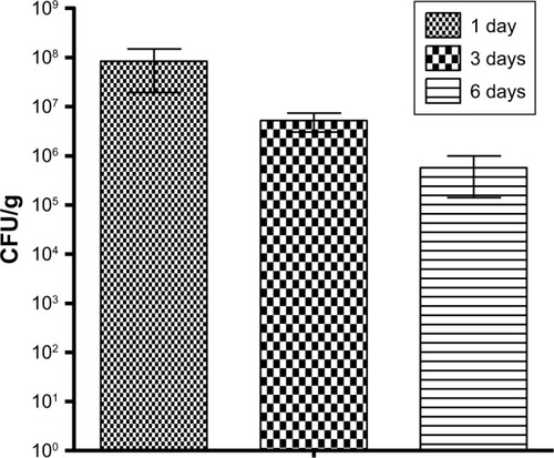
Figure 6 CFU counts in mammary glands after treatment.
Note: The horizontal line indicates the CFU detection limit.
Abbreviations: CFU, colony forming unit; IC, infected control group and treated with 0.9% [w/v] NaCl solution; SLN, solid lipid nanoparticle.
![Figure 6 CFU counts in mammary glands after treatment.Note: The horizontal line indicates the CFU detection limit.Abbreviations: CFU, colony forming unit; IC, infected control group and treated with 0.9% [w/v] NaCl solution; SLN, solid lipid nanoparticle.](/cms/asset/ecce6aa8-0b52-44aa-9126-032c346ef16e/dijn_a_12194135_f0006_b.jpg)