Figures & data
Figure 1 Chemical structure of the bioinspired hydrogels.
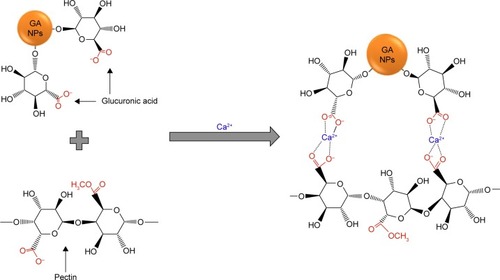
Figure 2 Morphological characterization of the bioinspired hydrogels.
Notes: AFM image (A) and DLS analysis (B) of the nanoparticles from gum arabic, the scale bar=500 nm. The nanostructure (network) investigated from the bioinspired hydrogels (C); the scale bar=500 nm.
Abbreviations: AFM, atomic force microscopy; DLS, dynamic light scattering.
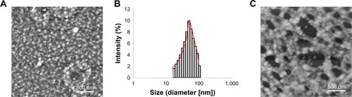
Figure 3 The bFGF release profiles from the bioinspired hydrogels (n=3).
Abbreviation: bFGF, basic fibroblast growth factor.
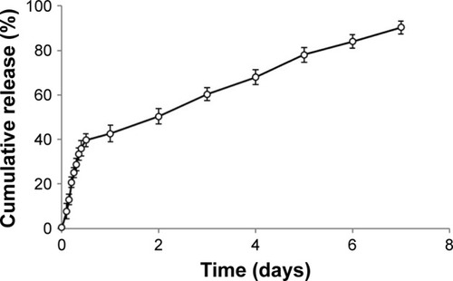
Figure 4 The dermal fibroblast cell proliferation on the bioinspired hydrogels with and without bFGF, negative control (non-treated), and positive control (commercial product; n=3).
Note: Significant differences between samples means are indicated; *P<0.05.
Abbreviation: bFGF, basic fibroblast growth factor.
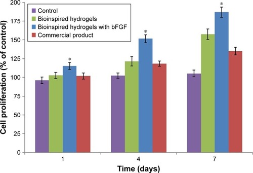
Figure 5 The in vitro scratch assays of L929 dermal fibroblast cells treated with control, bioinspired hydrogels with and without bFGF, and commercial product.
Notes: The images (A) and the analyzed results (B) showed the extent of wound closure in scratch assays after 12 and 24 hours for different groups. Error bars indicate SD. Significant differences between samples means are indicated; *P<0.05.
Abbreviation: bFGF, basic fibroblast growth factor.

Figure 6 The images (A) and the analyzed results (B) of in vivo wound closure studies for control, bioinspired hydrogels with and without bFGF, and commercial product. Error bars indicate SD.
Note: Significant differences between samples means are indicated; *P<0.05.
Abbreviation: bFGF, basic fibroblast growth factor.
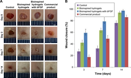
Figure 7 Histological images of H&E (A) and Masson’s trichrome (B) stained sections after 14 days of wound healing.
Abbreviation: bFGF, basic fibroblast growth factor.
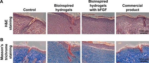
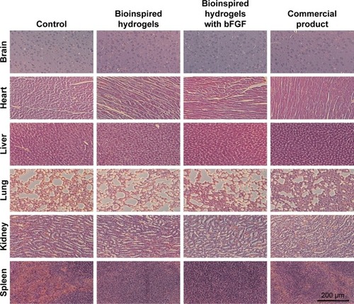
Figure 8 H&E stained organs revealed no significant signs of toxicity.
Abbreviation: bFGF, basic fibroblast growth factor.