Figures & data
Table 1 Generally used nanomedicines for antimicrobial agents
Figure 1 Schematic representation of the development of various nanostructures and in vitro and in vivo evaluation.
Abbreviations: DSC, differential scanning calorimeter; DLS, diffraction light scattering; ELS, electrophoretic light scattering; AFM, atomic force microscopy; XRD, X-ray diffraction; IVIVC, in vitro in vivo correlation; SLN, solid lipid nanoparticle; NLC, nanostructured lipid carrier; SNEDDS, self-nanoemulsifying drug delivery systems; SEM, scanning electron microscopy; TEM, transmission electron microscopy.
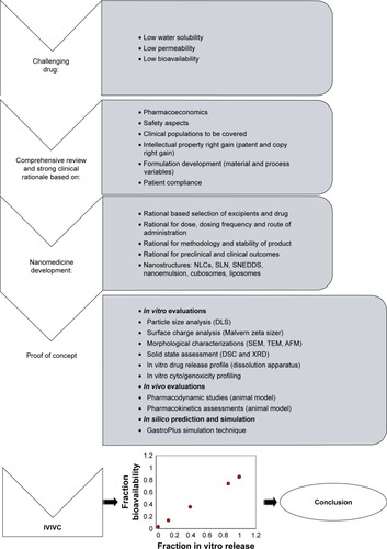
Figure 2 Schematic illustration of the possible mechanisms involved in microbial killing using nanomedicines (nanocarriers) composed of lipids and surfactants. Possible mechanisms are: (A) lipid–lipid interactions, (B) cationic nanomedicine interactions with negatively charged bacterial surfaces, (C) detergency-like action, (D) cytoplasmic content oozing out through developed tiny pores, (E) micelles loaded with extracted cellular lipids, (F) accumulated excipients to toxic levels to produce detrimental effects, and (G) P-gp efflux pump inhibition by specific excipients (reported labrasol, tween 80) and drugs (ketoconazole).Citation4,Citation19,Citation31,Citation67
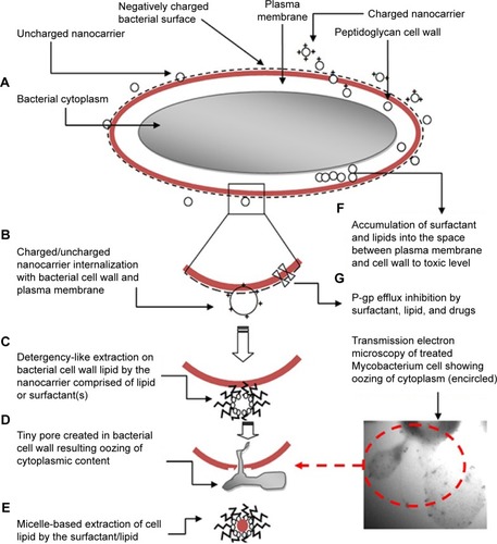
Figure 3 SEM microphotograph depicting the lethal effect of nanoemulsions loaded with ethambutol against Mycobacterium smegmatis: (A) control untreated, (B) damaged cells when treated with ethambutol-loaded nanoemulsion, (C and D) chemical structure of high molecular weight amphotericin B with cyclic hydrocarbon chain (amphiphilic molecule) with three-dimensional presentation, (E) rhodamine-123-probed nanoemulsion and dye-free AmB nanoemulsion, and (F) pseudoternary phase diagram for AmB-loaded nanoemulsion (Smix: 1:3).
Abbreviations: SEM, scanning electron microscopy; AmB, amphotericin B.
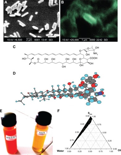
Table 2 A short review of nanomedicines for the delivery of antimicrobial agents
Table 3 List of common components employed in nanomedicines
Table 4 Reports suggesting anti-Mycobacterium activity of certain lipids or fatty acids
Figure 4 Results of the experimental design-based optimization of nanoemulsions comprised of labrasol and capmul MCM C8 with respective ZOI (zone of inhibitions) against MS-942 and MS-995 strains (Mycobacterium smegmatis). A–C are three-dimensional surface response plots, interaction curves and residual plots of ZOI against MS-942, respectively. Similarly, D–F are three-dimensional surface response plots, interaction curves, and residual plots of ZOI against MS-995, respectively.
Abbreviation: ZOI, zone of inhibition.
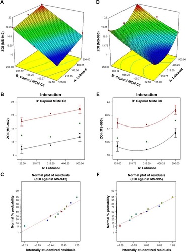
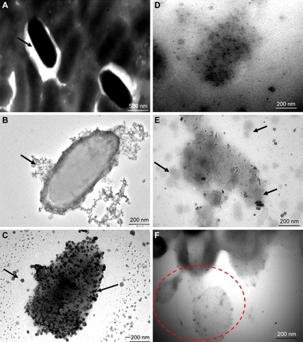
Figure 5 Results of a TEM study on the morphological assessment when treated with optimization placebo nanoemulsions comprised of labrasol and capmul MCM C8 in comparison with untreated tubercular bacilli (Mycobacterium tuberculosis H37 Rv). These representative images portrayed sequential events after 30 minutes of treatment with explored nanoemulsions, suggesting a possible mechanistic view of action against a tubercular strain. (A) Normal intact smooth margin of tubercular bacilli, (B and C) progressive damage of a bacterial cell wall followed by fragmentation (black arrows), (D) loss of integrity of bacterial cell wall, (E) mechanistic view of nanoemulsion action after adherence around the bacterial cell surface (nano globules around the surface), and (F) oozing out of the cytoplasmic content of bacterium after nanoemulsion mediated cell wall damage.
Abbreviation: TEM, transmission electron microscopy.