Figures & data
Figure 1 Scheme of conjugated paclitaxel to MWNT-g-PCA.
Abbreviations: MWNT, multiwalled carbon nanotube; PCA, poly citric acid.
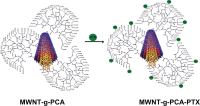
Figure 2 Differential scanning calorimetry thermograms of A) MWNT-g-PCA, B) paclitaxel, C) physical mixture of MWNT-g-PCA and paclitaxel (1:1 w/w), and D) MWNT-g-PCA-PTX conjugates containing 40% w/w paclitaxel.
Abbreviations: MWNT, multiwalled carbon nanotube; PCA, poly citric acid; PTX, paclitaxel.
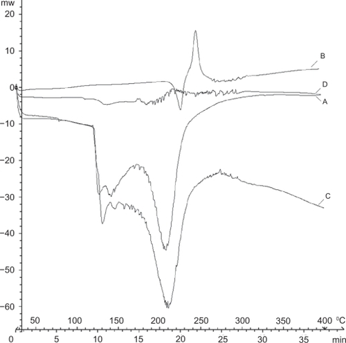
Figure 3 Atomic force microscopy images of A) oxidized MWNTs, B–D) MWNT-g-PCA and E–G) MWNT-g-PCA-PTX conjugates on mica surface. A, B, and E are topographic and C, D, F, and G are phase contrast images.
Abbreviations: MWNT, multiwalled carbon nanotube; PCA, poly citric acid; PTX, paclitaxel.
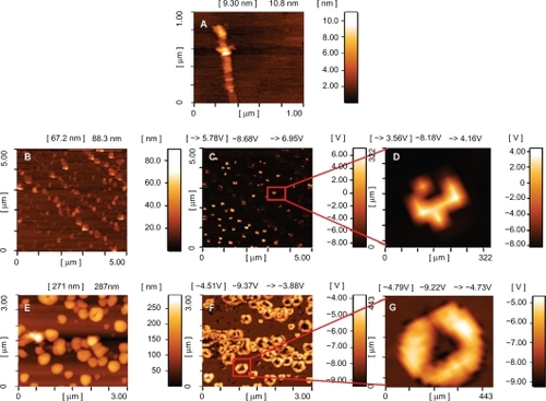
Figure 4 Transmission electron microscopy images of MWNT-g-PCA-PTX conjugates.
Abbreviations: MWNT, multiwalled carbon nanotube; PCA, poly citric acid; PTX, paclitaxel.
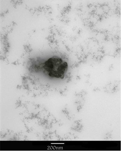
Figure 5 Release profiles of paclitaxel from MWNT-g-PCA-PTX conjugates at preselected time intervals. Data are presented as means ± standard deviations (n = 3).
Abbreviations: MWNT, multiwalled carbon nanotubes; PCA, poly citric acid; PTX, paclitaxel.
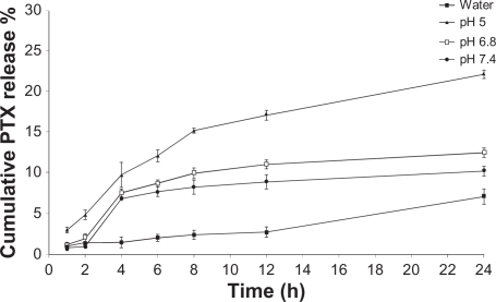
Table 1 IC50s (nM) of paclitaxel and conjugated MWNT-g-PCA-PTX against A549 and SKOV3 cell lines at different incubation times
Figure 6 Cytotoxicity of conjugated MWNT-g-PCA, MWNT-g-PCA-PTX, and paclitaxel against A549 (A) and SKOV3 (B) cells at 5, 10, 20, and 40 nM drug concentrations. Incubation time was 24 hours and 48 hours. Data are presented as means ± standard deviations (n = 3).
Abbreviations: MWNT, multiwalled carbon nanotube; PCA, poly citric acid; PTX, paclitaxel.
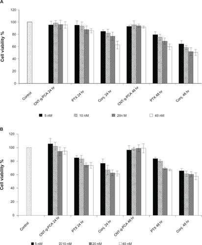
Figure 7 Cytotoxicity of MWNT-g-PCA, MWNT-g-PCA-PTX, and paclitaxel against A549 (A, B) and SKOV3 (C, D) cells. Incubation time was 96 hours in A and C, and 120 hours in B and D. Data are presented as means ± standard deviations (n = 3).
Abbreviations: MWNT, multiwalled carbon nanotubes; PCA, poly citric acid; PTX, paclitaxel.
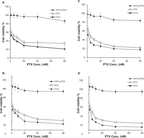
Figure S1 Infrared spectra of the A) oxidized MWNTs, B) MWNT-g-PCA, C) PTX, and D) MWNT-g-PCA-PTX conjugates containing 40% w/w PTX.
Abbreviations: MWNT, multiwalled carbon nanotube; PCA, poly citric acid; PTX, paclitaxel.
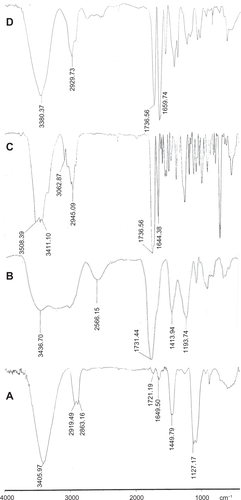
Figure S2 1H nuclear magnetic resonance spectra of A) paclitaxel, B) MWNT-g-PCA, and C) MWNT-g-PCA-PTX conjugates containing 40% w/w paclitaxel.
Abbreviations: MWNT, multiwalled carbon nanotube; PCA, poly citric acid; PTX, paclitaxel.
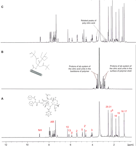
Figure S3 13C nuclear magnetic resonance spectra of A) PTX, B) MWNT-g-PCA, and C) MWNT-g-PCA-PTX conjugates containing 40% w/w paclitaxel.
Abbreviations: MWNT, multiwalled carbon nanotube; PCA, poly citric acid; PTX, paclitaxel.
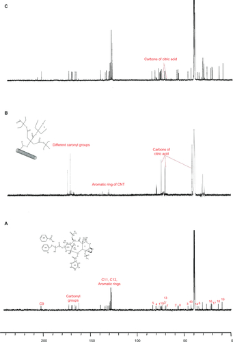
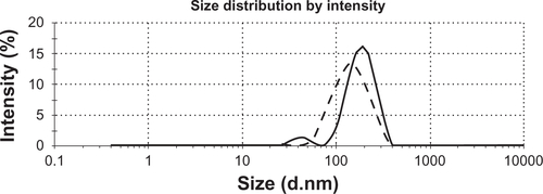
Figure S4 Dynamic light scattering analysis of MWNT-g-PCA (dotted line) and MWNT-g-PCA-PTX conjugate (solid line).
Abbreviations: MWNT, multiwalled carbon nanotubes; PCA, poly citric acid; PTX, paclitaxel.