Figures & data
Table 1 PD-L1 mAb content of the PEG-PCL-mAb NPs of various NPs/mAb ratios used in the process
Figure 1 Synthesis of DOC-PEG-PCL-mAb NPs and XPS spectrum results.
Notes: (A) Schematic illustrating the fabrication of PD-L1 mAb-conjugated PEG-PCL NPs: the NPs comprise a PCL core with DOC loaded, a hydrophilic and stealth PEG shell on the surface of the core, and a mAb coating. (B) Representative XPS spectrum and N 1s peak of different NPs groups.
Abbreviations: DOC, docetaxel; mAb, monoclonal antibody; NP, nanoparticle; PEG-PCL, poly (ethylene glycol)-poly (ε-caprolactone); XPS, X-ray photoelectron spectroscopy.
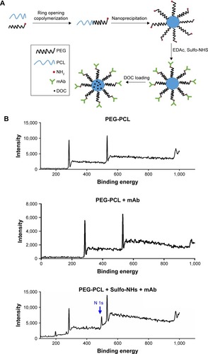
Table 2 Comparison of the characteristics of PEG-PCL, DOC-PEC-PCL-IgG, and DOC-PEG-PCL-mAbs
Figure 2 The structural characterization and surface morphology of the NPs.
Notes: (A) The size distribution of the DOC-PEG-PCL-mAb NPs determined by DLS. (B) Stability of the DOC-PEG-PCL-mAb NPs. The diameter of the NPs was determined by DLS, and each value represents the mean±SD. (C) Representative FESEM images of the DOC-PEG-PCL-mAb NPs. Scale bar represents 500 nm and 1 µm.
Abbreviations: DLS, dynamic light scattering; DOC, docetaxel; FESEM, field emission scanning electron microscopy; mAb, monoclonal antibody; NP, nanoparticle; PEG-PCL, poly (ethylene glycol)-poly (ε-caprolactone).
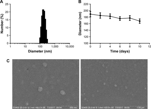
Figure 3 In vitro drug release profiles of the DOC-PEG-PCL-mAb NPs in pH 7.4 PBS buffer at 37°C where the measurement was made from 1 hour to 10 days.
Note: The data are represented as the mean±SEM, n=3.
Abbreviations: DOC, docetaxel; mAb, monoclonal antibody; NP, nanoparticle; PEG-PCL, poly (ethylene glycol)-poly (ε-caprolactone); SEM, standard error of the mean.
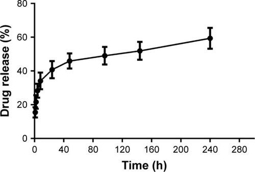
Figure 4 Cellular uptake analyses in MKN45 cells.
Notes: The hydrophobic Coumarin 6 was used to mimic the existence of docetaxel. The same concentration of well-dispersed fluorescent NPs (0.125 mg/mL) and Coumarin 6-PEG-PCL-mAb NPs was applied for incubation with the PD-L1 plasmid transfected and untransfected MKN45 cells for 1 hour (A) and 2 hours (B) at 37°C. Scale bars are labeled on the figures. **P<0.01, and *P<0.05 were considered significant.
Abbreviations: mAb, monoclonal antibody; NP, nanoparticle; PD-L1, programmed death-ligand 1; PEG-PCL, poly (ethylene glycol)-poly (ε-caprolactone).
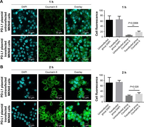
Figure 5 In vitro viability of three types of gastric cancer cells treated with blank NPs, PEG-PCL, PEG-PCL-IgG, PEG-PCL-mAb, free DOC, DOC-PEG-PCL, DOC-PEG-PCL-IgG, and DOC-PEG-PCL-mAb NPs at different drug concentrations after 48 hours of incubation.
Notes: (A) PD-L1 expression level of MGC803, MKN45, and HGC27 cells before and after PD-L1 plasmid transfection by flow cytometry and Western blot analysis. (B) In vitro viability of three kinds of gastric cancer cells. Data were expressed as the mean±SD of three independent experiments. ***P<0.005, **P<0.01, and *P<0.05 were considered significant.
Abbreviations: APC, allophycocyanin; DOC, docetaxel; IgG, Immunoglobulin G; mAb, monoclonal antibody; NP, nanoparticle; PD-L1, programmed death-ligand 1; PEG-PCL, poly (ethylene glycol)-poly (ε-caprolactone).
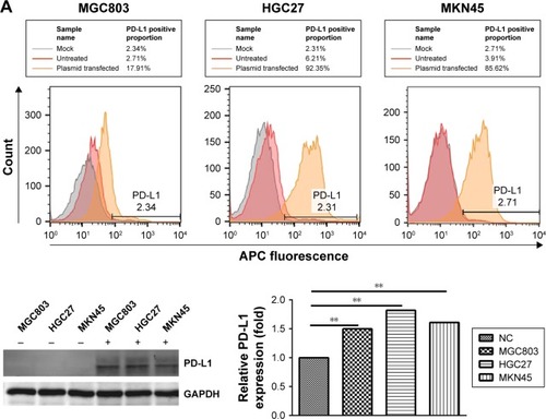
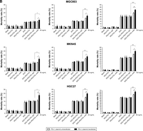
Figure 6 Apoptosis of HGC27 cells after exposure to blank PEG-PCL, PEG-PCL-IgG, PEG-PCL-mAb, DOC, DOC-PEG-PCL-IgG, and DOC-PEG-PCL-mAb NPs.
Notes: (A) Scatter plot indicating the cell populations in apoptotic and necrotic quadrants with the percentage of cancer cell death posttreatment of different NPs. (B) Western blot data of apoptosis showing the expression of proapoptotic and antiapoptotic proteins. Data were presented as the mean±SD of three independent experiments. ***P<0.005, **P<0.01, and *P<0.05 were considered significant.
Abbreviations: Bcl-2, B-cell lymphoma 2; DOC, docetaxel; IgG, immunoglobulin G; mAb, monoclonal antibody; NP, nanoparticle; PEG-PCL, poly (ethylene glycol)-poly (ε-caprolactone).
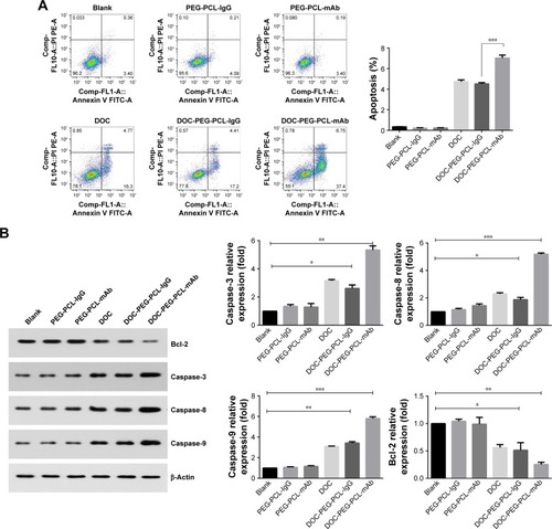
Figure 7 DOC-PEG-PCL-mAb NPs enhance G2/M arrest.
Notes: (A) The influence of cell cycle was analyzed using PI/RNase buffer. Compared with control group, DOC-PEG-PCL-mAb NPs significantly induced cell cycle arrest at the G2/M phase. (B) Cyclin A and B protein accumulated in the DOC-PEG-PCL-mAb NPs-treated group. ***P<0.005 and **P<0.01 were considered significant.
Abbreviations: DOC, docetaxel; IgG, immunoglobulin G; mAb, monoclonal antibody; NP, nanoparticle; PE-A, phycoerythrin-A; PEG-PCL, poly (ethylene glycol)-poly (ε-caprolactone); PI, propidium iodide.
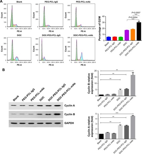
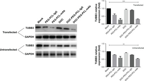
Figure 8 DOC-PEG-PCL-mAb NPs influence microtubule dynamics.
Notes: An evident reduction of TUBB3 expression was observed after treatment in the mAb-labeled NPs groups compared with other groups. ***P<0.005 and **P<0.01 were considered significant.
Abbreviations: DOC, docetaxel; GAPDH, glyceraldehyde-3-phosphate dehydrogenase; IgG, immunoglobulin G; mAb, monoclonal antibody; NP, nanoparticle; PEG-PCL, poly (ethylene glycol)-poly (ε-caprolactone); TUBB3, tubulin beta-3k.