Figures & data
Figure 1 (A) Microscopic section of the Ag-nHA-nTiO2/PA66 (AM) membrane (H&E ×100); (B) microscopic section of the nHA/PA66 membrane (H&E ×100); (C) microscopic section of the e-PTFE membrane (H&E ×200).
Notes: +, the observe; –, the reverse.
Abbreviations: e-PTFE, expanded poly tetrafluroethylene; H&E, hematoxylin and eosin stain; PA66, polyamide-66.
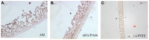
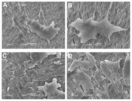
Figure 2 (A) SEM image of surface of obverse of the Ag-nHA-nTiO2/PA66 membrane (×500); (B) SEM image of surface of reverse of the Ag-nHA-nTiO2/PA66 membrane (×500); (C) SEM image of surface of obverse of the e-PTFE membrane (×2000); (D) SEM image of surface of reverse of the e-PTFE membrane (×2000).
Abbreviations: e-PTFE, expanded poly tetrafluroethylene; PA66, polyamide-66; SEM, scanning electronic microscope.

Table 1 MTT values of experimental and control groups at different times
Table 2 Mean CVA values of MG63 osteoblast-like cells on experimental and control groups
Table 3 Alkaline phosphatase activity at 1, 3, 5, and 7 days in experimental and control groups
Table 4 Ca2+ concentration at 1, 3, 5, and 7 days in experimental and control groups
Figure 3 (A and B) Adhesion of MG63 osteoblast-like cells on day 1 on the Ag- nHA-nTiO2/PA66 membrane (SEM * 400)/(SEM * 1000). (C and D) Adhesion of MG63 osteoblast-like cells on day 5 day on the Ag-nHA-nTiO2/PA66 membrane (SEM * 400)/(SEM * 1000).
Abbreviations: PA66, polyamide-66; SEM, scanning electronic microscope.
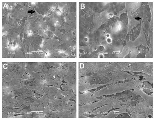
Figure 4 (A and B) Adhesion of MG63 osteoblast-like cells on day 1 on the nHA/PA66 membrane (SEM * 400)/(SEM * 1000). (C and D) Adhesion of MG63 osteoblast-like cells on day 5 on the nHA/PA66 membrane (SEM * 400)/(SEM * 1000).
Abbreviations: PA66, polyamide-66; SEM, scanning electronic microscope.
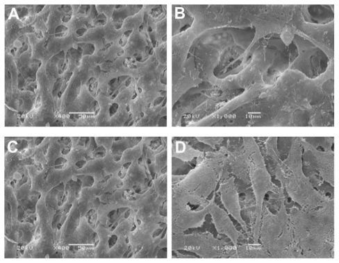
Figure 5 (A and B) Adhesion of MG63 osteoblast-like cells on day 1 on the e-PTFE membrane (SEM * 400)/(SEM * 1000). (C and D) Attachment of MG63 osteoblastlike cells on day 5 day on the e-PTFE membrane (SEM * 400)/(SEM * 1000).
Abbreviations: e-PTFE, expanded poly tetrafluroethylene; SEM, scanning electronic microscope.