Figures & data
Figure 1 Characterization of SeNPs and Se@RBV.
Notes: (A) TEM images of SeNPs (a) and Se@RBV (b). (B) Elemental composition analysis of Se@RBV by EDX. (C) Size distributions of SeNPs and Se@RBV. (D) Zeta potentials of SeNPs and Se@RBV.
Abbreviations: EDX, energy-dispersive X-ray spectroscopy; RBV, ribavirin; SeNPs, selenium nanoparticles; Se@RBV, SeNPs loaded with RBV; TEM, transmission electron microscopy.
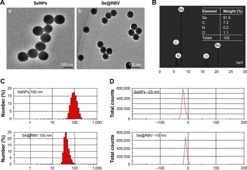
Figure 2 Cell viability and viral proliferation.
Notes: MDCK cells infected with H1N1 virus were untreated (Mock) or treated with RBV, SeNPs, and Se@RBV for 48 hours. Cells without infection and treatment were performed as control. Cell viability (A) and virus titer of the culture supernatant (B) were detected. Cytopathic effect was observed under a microscope (C). *P<0.05.
Abbreviations: MDCK, Madin-Darby Canine Kidney; RBV, ribavirin; SeNPs, selenium nanoparticles; Se@RBV, SeNPs loaded with RBV.
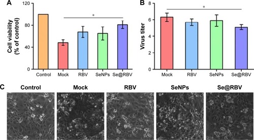
Figure 3 Intracellular localization of Se@RBV.
Notes: MDCK cells were treated with coumarin-6-loaded Se@RBV for 90 minutes and stained with lyso tracker for lysosome and DAPI for nucleus. Cells were observed under a fluorescent microscope at a different time.
Abbreviations: DAPI, 4′6-diamidino-2-phenyindole; MDCK, Madin-Darby Canine Kidney; RBV, ribavirin; SeNPs, selenium nanoparticles; Se@RBV, SeNPs loaded with RBV.
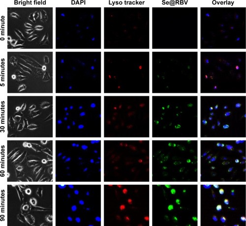
Figure 4 Restriction on caspase-3 activation in vitro.
Notes: MDCK cells infected with H1N1 virus were untreated (Mock) or treated with RBV, SeNPs, and Se@RBV for 48 hours. Cells without infection and treatment were performed as control. Caspase-3 activity was checked and expressions of proteins related to caspase-3 activation were detected. *P<0.05 and **P<0.01.
Abbreviations: MDCK, Madin-Darby Canine Kidney; RBV, ribavirin; SeNPs, selenium nanoparticles; Se@RBV, SeNPs loaded with RBV.
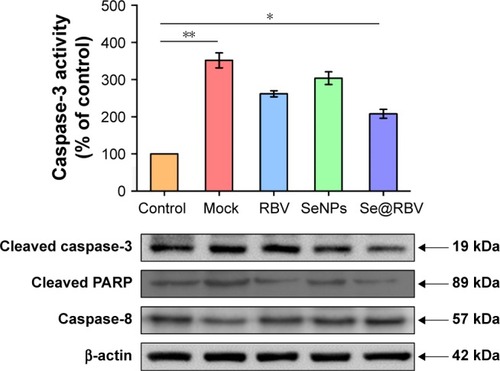
Figure 5 Inhibition of apoptosis signaling pathways in vitro.
Notes: MDCK cells infected with H1N1 virus were untreated (Mock) or treated with RBV, SeNPs, and Se@RBV for 48 hours. Cells without infection and treatment were performed as control. (A) Inhibition of apoptotic proteins. (B) Scheme of apoptosis signaling pathways.
Abbreviations: MDCK, Madin-Darby Canine Kidney; PARP, poly-ADP-ribose polymerase ; p-JNK, Jun-amino-terminal kinase; p38, phosphorylated 38; p53, phosphorylated 53; RBV, ribavirin; SeNPs, selenium nanoparticles; Se@RBV, SeNPs loaded with RBV.
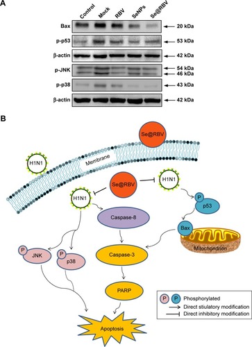
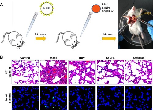
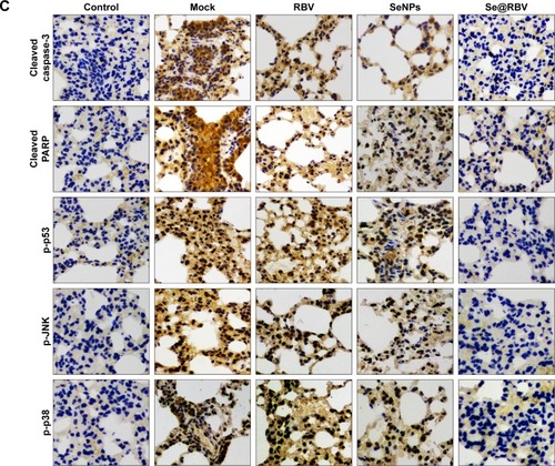
Figure 6 In vivo antiviral efficiency of Se@RBV.
Notes: Mice infected by H1N1 virus was treated with physiological saline (Mock), RBV, SeNPs, or Se@RBV (A). Mice without infection and treated with physiological saline were performed as control. H&E and tunel staining (B), as well as immunohistochemistry (C), was carried out.
Abbreviations: H&E, hematoxylin and eosin; MDCK, Madin-Darby Canine Kidney; PARP, poly-ADP-ribose polymerase; p-JNK, Jun-amino-terminal kinase; p38, phosphorylated 38; p53, phosphorylated 53; RBV, ribavirin; SeNPs, selenium nanoparticles; Se@RBV, SeNPs loaded with RBV.