
Figures & data
Figure 1 General structure of the glyconanotube used in this study.
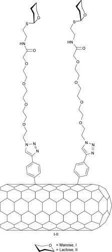
Figure 2 Synthetic pathway for the synthesis of derivatives 11 and 12.
Notes: (a) Boc2O, THF, 54%; (b) see ref Citation36; (c) CuSO4, sodium ascorbate, CH2Cl2/H2O, MW, 7 (62%), 8 (80%); (d) TFA, CH2Cl2, 9 (99%), 10 (80%); (e) MeONa/MeOH, 11 (79%), 12 (60%).
Abbreviations: THF, tetrahydrofuran; MW, microwave; TFA, trifluoroacetic acid.
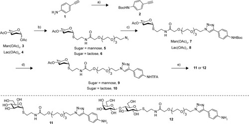
Figure 3 Synthetic pathway of derivatives I and II.
Notes: a refers to I: P2-SWCNT, oDCB/DMF; II: amyl nitrite, 65°C, 24 hours.
Abbreviations: SWCNT, single-walled carbon nanotube; oDCB, orthodichlorobenzene; DMF, dimethylformamide.
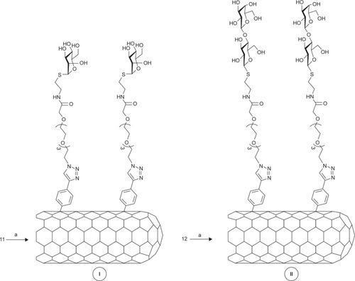
Table 1 Results on solubility/functionalization of derivatives glyconanotubes I and II
Figure 4 (A) UV/Vis/NIR spectra of long chain sugar-coated SWCNTs: SWCNTs (solid), I (red dotted line) and II (blue dashed line). (B) Comparative Raman spectra of SWCNTs (black), I (red), and II (blue) using a laser operating at 785 nm.
Abbreviations: UV/Vis/NIR, ultraviolet/visible/near infrared; SWCNTs, single-walled carbon nanotubes; ID, D-band intensity; IG, G-band intensity.
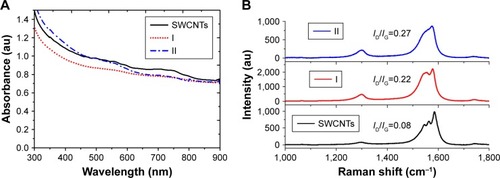
Figure 5 (A) TEM micrograph of water solutions of mannose-coated SWCNT I, (B) TEM micrograph of water solutions of lactose-coated SWCNT II, (C) STEM micrograph of water solutions of mannose-coated SWCNT I with HAADF detector, (D) STEM micrograph of water solutions of lactose-coated SWCNT II with HAADF detector, (E) SEM micrograph of water solutions of mannose-coated SWCNT I, and (F) SEM micrograph of water solutions of lactose-coated SWCNT II.
Abbreviations: TEM, transmission electronic microscopy; SWCNT, single-walled carbon nanotube; STEM, scanning transmission electronic microscopy; HAADF, high-angle darkfield; SEM, scanning electronic microscopy.
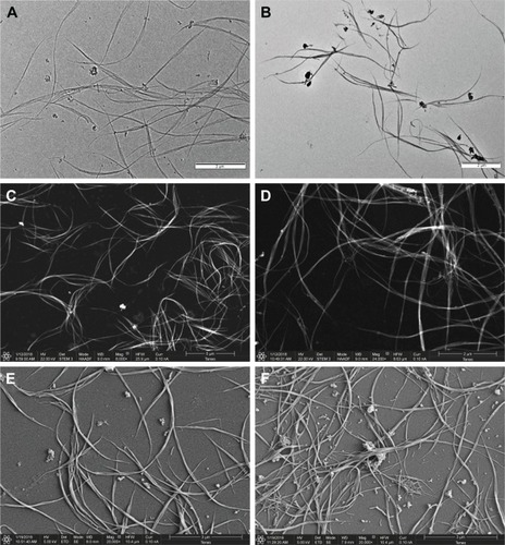
Figure 6 Thermograms by TGA of (A) mannose-coated SWCNT I (red) and (B) lactose-coated SWCNT II (blue), wherein compared the weight loss of the unfunctionalized SWCNTs (solid line), the starting carbohydrates (dotted), and the title compounds (dashed).
Abbreviations: TGA, thermogravimetric analysis; SWCNT, single-walled carbon nanotube.
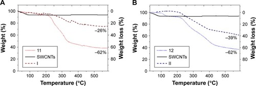
Figure 7 Schematic representation of the FimH adhesin promoted specific interaction of E. coli with mannose-coated SWCNTs I.
Abbreviations: SWCNTs, single-walled carbon nanotubes; E. coli, Escherichia coli.
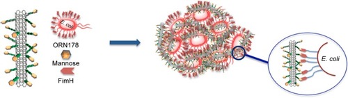
Figure 8 Representative fluorescence microscopy images of the interaction of glyconanotubes I and II with E. coli strain ORN178 and ORN208.
Notes: (A, B) E. coli strain ORN178 alone. (C, D) E. coli strain ORN208 alone. (E, F) Selective interaction of glyconanotube I with the E. coli strain ORN178. (G, H) Interaction of glyconanotube I with the E. coli strain ORN208. (I, J) Interaction of glyconanotube II with the E. coli strain ORN178. (K) CFU assay (see Figure S5 and for more information). The number of colonies formed in the absence (control) and presence of glyconanotube I for ORN178 and ORN208 cells is indicated. For conditions, see “Materials and methods” section in the text.
Abbreviations: SWCNTs, single-walled carbon nanotubes; E. coli, Escherichia coli; CFU, colony-forming unit.
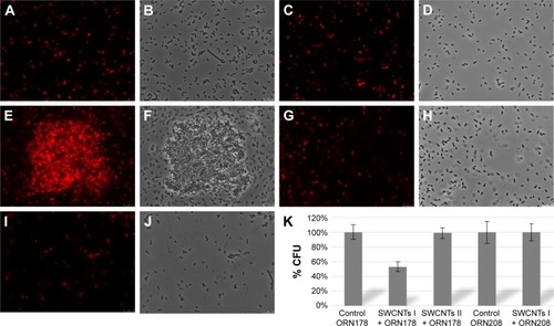
Figure 9 Representative, negatively stained TEM and sputter coated with gold, micrographs of the interaction of glyconanotubes I and II with E. coli strains ORN178 and ORN208.
Notes: (A) TEM micrograph of E. coli strain ORN178 alone. (B) TEM micrograph of E. coli strain ORN208 alone. (C) TEM micrograph of the lactose-coated glyconanotube II incubated with the E. coli strain ORN178. (D) SEM micrograph of sputter coated with gold of glyconanotube II incubated with E. coli strain ORN178. (E) TEM micrograph of the mannose-coated glyconanotube I incubated with the E. coli strain ORN178. (F) SEM micrograph of sputter coated with gold of glyconanotube I incubated with E. coli strain ORN178. For conditions, see “Materials and methods” section in the text. Size bars are shown.
Abbreviations: TEM, transmission electronic microscopy; SWCNTs, single-walled carbon nanotubes; E. coli, Escherichia coli; SEM, scanning electronic microscopy.
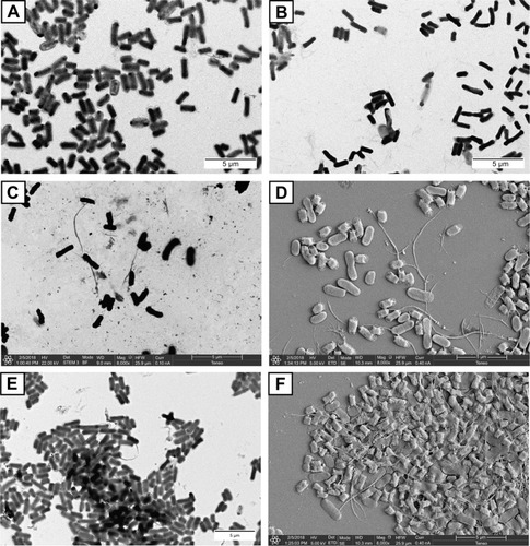
Figure S1 1H-NMR of final monomer 11.
Abbreviation: NMR, nuclear magnetic resonance.
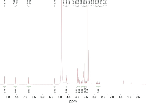
Figure S2 13C-NMR of final monomer 11.
Abbreviation: NMR, nuclear magnetic resonance.
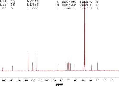
Figure S3 1H-NMR of final monomer 12.
Abbreviation: NMR, nuclear magnetic resonance.
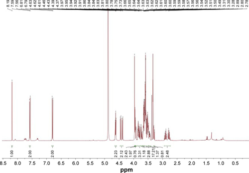
Figure S4 13C-NMR of final monomer 12.
Abbreviation: NMR, nuclear magnetic resonance.
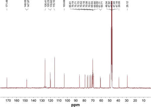
Figure S5 CFU reduction test of E. coli strains ORN178 and ORN208 with glyconanotubes I and II.
Abbreviations: CFU, colony forming units; E. coli, Escherichia coli.
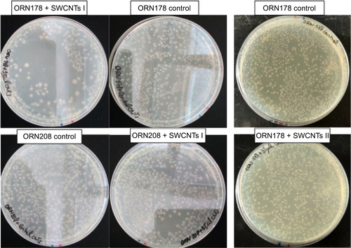
Figure S6 SEM image of bacterial agglutination by glyconanomaterial I.
Abbreviation: SEM, scanning electronic microscopy.
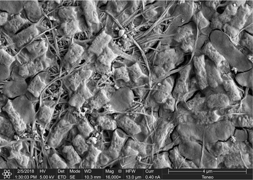
Figure S7 SEM image of no bacterial agglutination by glyconanomaterial II.
Abbreviation: SEM, scanning electronic microscopy.
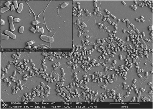
Table S1 CFU reduction test of E. coli strains ORN178 and ORN208 with glyconanotubes I and II