Figures & data
Figure 1 Fabrication of the NAC-loaded PLGA electrospun scaffold and PNNM electrospun scaffold.
Notes: Fabrication of the PLGA electrospun scaffold (A) showing fiber formation. Fibers are organized on the rotor receiver of the equipment. Gross view of the PNNM electrospun scaffold (B) NAC@MSN particles and NAC are embedded in the fibers. Reprinted from Acta Biomater, 8(5), Song B, Wu C, Chang J, Dual drug release from electrospun poly(lactic-co-glycolic acid)/mesoporous silica nanoparticles composite mats with distinct release profiles, 1901–1907, Copyright (2012), with permission from Elsevier.Citation22
Abbreviations: NAC, N-acetyl cysteine; PLGA, polylactic-co-glycolic acid; MSNs, mesoporous silica nanoparticles; PNNM, PLGA with free NAC and NAC@MSN.
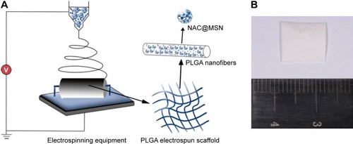
Table 1 Primer sequences for qRT-PCR
Figure 2 SEM image, TEM image, and particle size distribution of MSNs, and chemical analyses of MSNs after loading with NAC.
Notes: SEM image of MSNs (A). TEM image of MSNs (B). Particle size distribution of MSNs (C). Infrared spectra of MSNs and NAC@MSN (D). Arrows indicate the characteristic peaks of Si at 794, 995, and 1,095 cm−1, and the characteristic peaks of NAC at 1,392, 1,552, and 1,731 cm−1 (D). Thermogravimetric analysis of MSNs before and after loading with NAC (E). Derivative thermogravimetric analysis of MSNs before and after loading with NAC (F).
Abbreviations: SEM, scanning electron microscope; TEM, transmission electron microscopy; MSNs, mesoporous silica nanoparticles; NAC, N-acetyl cysteine; NAC@MSN, NAC-loaded MSN.
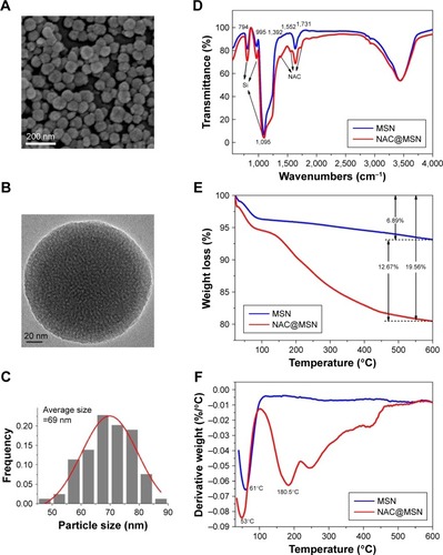
Figure 3 SEM image, TEM image, fiber diameter distribution, and degradation of pure PLGA, PN, PNM, and PNNM scaffolds.
Notes: SEM images, TEM images, fiber diameter distribution, and degradation after 21 days of pure PLGA scaffolds (A1–A5), PN scaffolds (B1–B5), PNM scaffolds (C1–C5), and PNNM scaffolds (D1–D5). The arrows point to MSNs (C3) and NAC@MSN (D3).
Abbreviations: SEM, scanning electron microscope; TEM, transmission electron microscopy; PLGA, polylactic-co-glycolic acid; PN, PLGA with free NAC; PNM, PLGA with NAC@MSN; PNNM, PLGA with free NAC and NAC@MSN; MSNs, mesoporous silica nanoparticles; NAC, N-acetyl cysteine; NAC@MSN, NAC-loaded MSN.
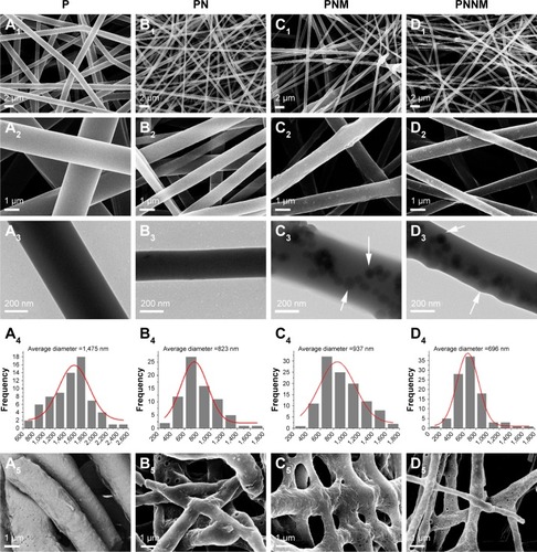
Figure 4 Water contact angle, swelling rate, nanoindentation hardness, and elastic modulus of pure PLGA, PN, PNM, and PNNM scaffolds.
Notes: Water contact angle of pure PLGA, PN, PNM, and PNNM scaffolds (A). Swelling rate of pure PLGA, PN, PNM, and PNNM scaffolds (B). Nanoindentation hardness and elastic modulus of pure PLGA, PN, PNM, and PNNM scaffolds (C and D). ** represents for P<0.01 and *** represents P<0.001 relative to the pure PLGA group (A); && represents P<0.01 and &&& represents P<0.001 relative to the PN group; ## represents P<0.01 relative to the PNM group.
Abbreviations: PLGA, polylactic-co-glycolic acid; PN, PLGA with free NAC; NAC, N-acetyl cysteine; PNM, PLGA with NAC@MSN; NAC@MSN, NAC-loaded MSNs; MSNs, mesoporous silica nanoparticles; PNNM, PLGA with free NAC and NAC@MSN.
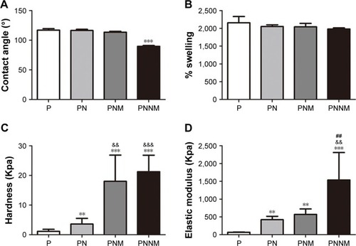
Figure 5 Drug-release kinetics of Si and NAC.
Notes: NAC release profiles of PN, PNM, and PNNM scaffolds during 0–280 hours in distilled water solution (A). Cumulative Si release profiles of PN, PNM, and PNNM scaffolds after 24 days in distilled water solution (B).
Abbreviations: PN, PLGA with free NAC; PNM, PLGA with NAC@MSN; PNNM, PLGA with free NAC and NAC@MSN; NAC, N-acetyl cysteine; NAC@MSN, NAC-loaded MSN.
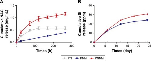
Figure 6 Cell viability and CLSM images of rBMSCs seeded on pure PLGA, PN, PNM, and PNNM scaffolds. CLSM images of shape of osteoblasts cultured under different conditions.
Notes: Cell viability values of rBMSCs (A) cultured on samples for 48 hours; Live/dead staining of rBMSCs (B1–E1) and actin cytoskeleton of rBMSCs (B2–E2) examined by CLSM at 48 hours on Pure PLGA (B1, B2), PN (C1, C2), PNM (D1, D2), and PNNM (E1, E2) sample. * represents P<0.05, ** represents P<0.01, and ***represents P<0.001 relative to the pure PLGA group (A). Green color indicates live rBMSCs in , red color indicates cytoskeleton of rBMSCs in B2–E2, and the blue color indicates cell nucleus in B2–E2. &&&represents P<0.001 relative to the PN group; ### represents P<0.001 relative to the PNM group.
Abbreviations: CLSM, confocal laser scanning microscopy; rBMSCs, rat bone marrow stromal cells; PN, PLGA with free NAC; PNM, PLGA with NAC@MSN; PNNM, PLGA with free NAC and NAC@MSN; PLGA, polylactic-co-glycolic acid; MSNs, mesoporous silica nanoparticles; NAC, N-acetyl cysteine; NAC@MSN, NAC-loaded MSN.
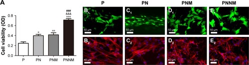
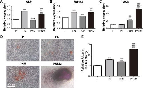
Figure 7 ALP, Runx2, and OCN expression, Alizarin red staining and relative qualification of Alizarin red.
Notes: ALP (A), Runx2 (B), and OCN (C) expression of rBMSCs seeded on pure PLGA, PN, PNM, and PNNM scaffolds for 7 days; Alizarin red staining (D) and relative qualification of Alizarin red (E) of rBMSCs cultured for 14 days with pure PLGA, PN, PNM, and PNNM scaffolds; ** represents P<0.01 and *** represents P<0.001 relative to the pure PLGA group; && represents P<0.01, &&& represents P<0.001 relative to the PN group; ### represents P<0.001 relative to the PNNM.
Abbreviations: ALP, alkaline phosphatase; OCN, osteocalcin; rBMSCs, rat bone marrow stromal cells; PN, PLGA with free NAC; PNM, PLGA with NAC@MSN; PNNM, PLGA with free NAC and NAC@MSN; PLGA, polylactic-co-glycolic acid; MSN, mesoporous silica nanoparticles; NAC, N-acetyl cysteine; NAC@MSN, NAC-loaded MSN.