Figures & data
Figure 1 Schematic representation of the Fe3O4@OA, Fe3O4@DMSA nanoparticles and Fe3O4@DMSA@Ab nanoprobes (A). Representative TEM imaging (B) and core diameter (C) analysis of the Fe3O4@OA (B(a), C(a)), Fe3O4@DMSA (B(b), C(b)) nanoparticles and Fe3O4@DMSA@Ab (B(c), C(c)) nanoprobes. Hydrodynamic size in number (D) and in volume (E) analysis of the Fe3O4@DMSA nanoparticles and Fe3O4@DMSA@Ab nanoprobes dispersed in water and stored at 4°C. The saturation magnetization (F) of the Fe3O4@DMSA nanoparticles and Fe3O4@DMSA@Ab nanoprobes.
Abbreviations: DMSA, 2,3-dimercaptosuccinic acid; TEM, transmission electron microscopy; TMAOH, tetramethylammonium hydroxide.
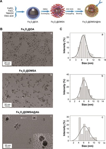
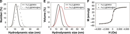
Figure 2 Schematic representation of Raji cells labeling with Fe3O4@DMSA@Ab nanoprobes and staining with Prussian blue for Fe (A). Detection of CD20 on the surface of Raji cells with a T/B kit and Fe3O4@DMSA@Ab (B, scale bar 100 µm). Control groups of Raji cells (B(a)) and K562 cells (B(d)). Detection of CD20 on Raji cells (B(b)). CD3 detecting on K562 cells (B(e)). Fe3O4@DMSA@Ab-labeled Raji cells (B(c)) and K562 cells (B(f)). TEM images of Raji (C(a, b)) and K562 (C(c, d)) cells incubated with Fe3O4@DMSA@Ab. MRI detection of Fe3O4@DMSA and Fe3O4@DMSA@Ab-labeled Raji cells (E) and K562 cells (F) and the corresponding 1/T2 variation as a function of [Fe] concentration (D).
Abbreviations: DMSA, 2,3-dimercaptosuccinic acid; TEM, transmission electron microscopy.
![Figure 2 Schematic representation of Raji cells labeling with Fe3O4@DMSA@Ab nanoprobes and staining with Prussian blue for Fe (A). Detection of CD20 on the surface of Raji cells with a T/B kit and Fe3O4@DMSA@Ab (B, scale bar 100 µm). Control groups of Raji cells (B(a)) and K562 cells (B(d)). Detection of CD20 on Raji cells (B(b)). CD3 detecting on K562 cells (B(e)). Fe3O4@DMSA@Ab-labeled Raji cells (B(c)) and K562 cells (B(f)). TEM images of Raji (C(a, b)) and K562 (C(c, d)) cells incubated with Fe3O4@DMSA@Ab. MRI detection of Fe3O4@DMSA and Fe3O4@DMSA@Ab-labeled Raji cells (E) and K562 cells (F) and the corresponding 1/T2 variation as a function of [Fe] concentration (D).Abbreviations: DMSA, 2,3-dimercaptosuccinic acid; TEM, transmission electron microscopy.](/cms/asset/4762264c-c5f5-4fc3-91bf-30de2eee0399/dijn_a_12190654_f0002_c.jpg)
![Figure 2 Schematic representation of Raji cells labeling with Fe3O4@DMSA@Ab nanoprobes and staining with Prussian blue for Fe (A). Detection of CD20 on the surface of Raji cells with a T/B kit and Fe3O4@DMSA@Ab (B, scale bar 100 µm). Control groups of Raji cells (B(a)) and K562 cells (B(d)). Detection of CD20 on Raji cells (B(b)). CD3 detecting on K562 cells (B(e)). Fe3O4@DMSA@Ab-labeled Raji cells (B(c)) and K562 cells (B(f)). TEM images of Raji (C(a, b)) and K562 (C(c, d)) cells incubated with Fe3O4@DMSA@Ab. MRI detection of Fe3O4@DMSA and Fe3O4@DMSA@Ab-labeled Raji cells (E) and K562 cells (F) and the corresponding 1/T2 variation as a function of [Fe] concentration (D).Abbreviations: DMSA, 2,3-dimercaptosuccinic acid; TEM, transmission electron microscopy.](/cms/asset/2d940c6f-3dd9-42b2-b0e6-5535e69a260c/dijn_a_12190654_f0002a_c.jpg)
Figure 3 Viability and apoptosis of Raji and K562 cells after culturing with rituximab, Fe3O4@DMSA, and Fe3O4@DMSA@Ab for 24 hours, detected by CCK-8 (A,B), AO/EB fluorescence staining (C, Scale bar 100 µm), and FlowCytometry (D) of Raji and K562 cells after 24 h.
Abbreviations: DMSA, 2,3-dimercaptosuccinic acid; CCK-8, cell counting kit-8.

Figure 4 Schematic illustration of the possible mechanism of Fe3O4@DMSA@Ab-mediated apoptosis in Raji cells (A). The intracellular Ca2+ flux of Raji cells (B) and K562 cells (C) after incubation with rituximab, Fe3O4@DMSA, and Fe3O4@DMSA@Ab for 24 hours. Measurement of corresponding intracellular ROS (D) and mitochondrial membrane potential (ΔΨm) (F) in cells. Apoptosis evaluation of Raji cells after incubating with or without rituximab, rituximab and Fe3O4@DMSA together, and Fe3O4@ DMSA@Ab (E).
Abbreviation: DMSA, 2,3-dimercaptosuccinic acid.
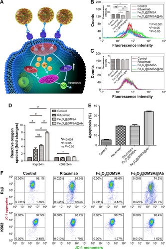
Figure 5 Expression of Bcl-2 and Bax protein after treatment of Raji and K562 cells with rituximab, Fe3O4@DMSA, and Fe3O4@DMSA@Ab (A), and the corresponding changes in Bcl-2/β-actin and Bax/β-actin (B).
Abbreviation: DMSA, 2,3-dimercaptosuccinic acid.

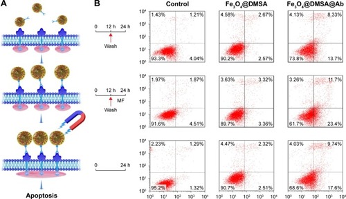
Figure 6 Schematic representation of CD20 clusters induced by Fe3O4@DMSA@Ab in the presence of a static magnetic field (A). Apoptosis of Raji cells detected by flow cytometry (B), Raji cells treated with Fe3O4@DMSA in the presence and absence of MF, Raji cells treated with Fe3O4@DMSA@Ab in the presence and absence of MF, and Raji cells treated with Fe3O4@DMSA@Ab for 24 hours.
Abbreviations: DMSA, 2,3-dimercaptosuccinic acid; FACS, FlowCytometry; MF, magnetostatic field.