Figures & data
Table 1 The effect of concentration on the characteristics of Heparin/Chitosan nanoparticles
Table 2 The effect of pH value on the characteristics of Heparin/Chitosan nanoparticles
Figure 1 The morphology of scaffolds. A) Morphology of photo-oxidative cross-linked decellularized scaffolds (SF-DP) from bovine jugular vein (BJV), magnification × 10,000. B) Morphology of SF-DP, magnification × 30,000. C) Morphology of heparin/chitosan (HEP/CS) nanoparticle-immobilized scaffold (SF-NP), magnification × 10,000. D) Morphology of HEP/CS nanoparticle-immobilized scaffold (SF-NP), magnification × 30,000. E) Surface roughness of SF-DP determined by atomic force microscopy (AFM). F) Surface roughness of SF-NP determined by AFM.
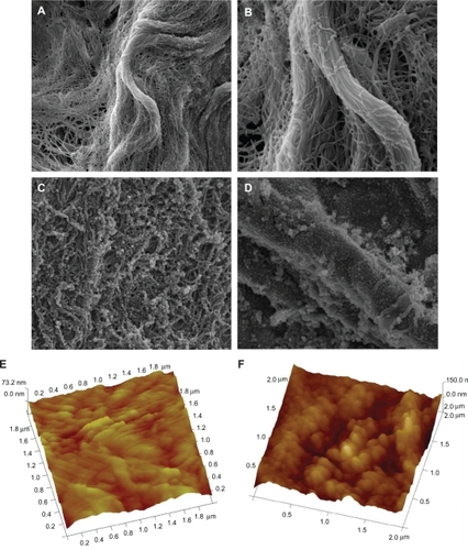
Figure 2 The characteristics of nanoparticle (NP)-delivered vascular endothelial growth factor (VEGF). A) Entrapping of VEGF in a concentration-dependent manner. B) Controlled release of VEGF from NPs localized at scaffolds. In the EDC-modified SF-NP, massive NPs entrapping VEGF still existed on the surface of scaffolds 4 weeks (C) and 10 weeks (D).
Abbreviations: EDC, 1-(3-Dimethylaminopropyl)-3-ethylcarbodiimide hydrochloride; DP, decellularized scaffolds; SF, scaffolds; V/VEGF, vascular endothelial growth factor
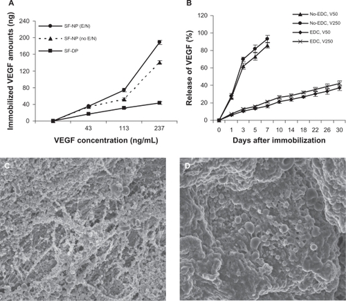
Figure 3 Nanoparticle-delivered VEGF stimulates endothelial cell proliferation and tube formation. A) MTT assay of the viability and proliferation of endothelial cell (EC) on scaffolds. B) Morphological changes of EC at 2, 3, or 7 days after incubation. SF-NP-VEGF showed more elongated EC cells and circular structures (tube formation) at day 3 and day 7.
Abbreviations: DP, decellularized scaffolds; MTT, 3-(4,5-dimethylthiazol-2-yl)2,5-diphenyltetrazolium bromide; NP, nanoparticles; SF, scaffolds; V/VEGF, vascular endothelial growth factor
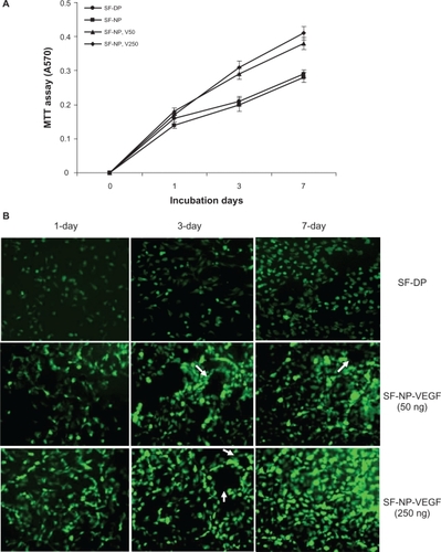
Figure 4 New vessel formation in SF-NP-VEGF implants. More new vessels were clearly seen in SF-NP-VEGF scaffolds at 4 weeks and 8 weeks after implantation.
Abbreviations: DP, decellularized scaffolds; NP, nanoparticle; SF, scaffolds; VEGF, vascular endothelial growth factor
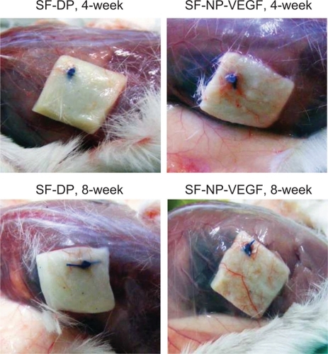
Figure 5 H&E staining of implant sections and capillary density. A) H&E staining showed new capillaries in repopulated layer and tissues surrounding the implants at 4 and 8 weeks, original magnification × 200. B) The capillary density calculated as the number of capillaries per mm2.
Notes: N = 8; P < 0.001.
Abbreviations: H&E, hematoxylin and eosin; DP, decellularized scaffolds; NP, nanoparticle; SF, scaffolds; VEGF, vascular endothelial growth factor
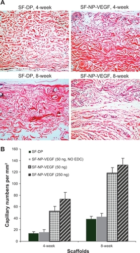
Figure 6 CD31staining of implant sections. The endothelial cells were stained by anti-CD31 antibody, original magnification × 200.
Note: The arrows indicate the positive staining.
Abbreviations: CD31, cluster of differentiation molecule 31; DP, decellularized scaffolds; NP, nanoparticle; SF, scaffolds; VEGF, vascular endothelial growth factor
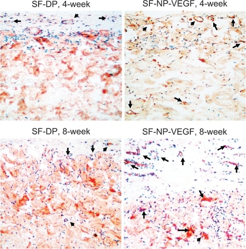
Figure 7 Immunohistochemical staining of fibroblasts. The fibroblasts were stained by anti-vimentin antibody, original magnification × 200. The red stained cells are the fibroblasts.
Note: The arrows indicate positive staining.
Abbreviations: DP, decellularized scaffolds; NP, nanoparticle; SF, scaffolds; VEGF, vascular endothelial growth factor
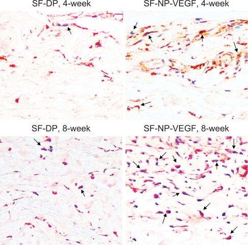
Figure 8 Immunohistochemical staining of macrophages. The macrophages were stained by anti-CD68 antibody, original magnification × 100.
Note: The arrows indicated red staining cells are macrophages.
Abbreviations: CD68, cluster of differentiation molecule 68; DP, decellularized scaffolds; NP, nanoparticle; SF, scaffolds; VEGF, vascular endothelial growth factor
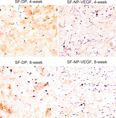
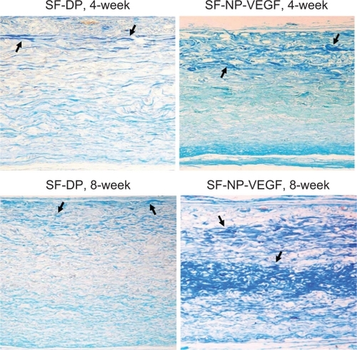
Figure 9 Herovici staining of collagen fibrils. Staining showed extracellular matrix components of scaffolds at 4 and 8 weeks implantation, original magnification × 200.
Notes: Red indicates the mature collagen fibrils and blue indicates new collagen fibrils. The arrows indicate positive staining of new collagen fibrils.
Abbreviations: DP, decellularized scaffolds; NP, nanoparticle; SF, scaffolds; VEGF, vascular endothelial growth factor
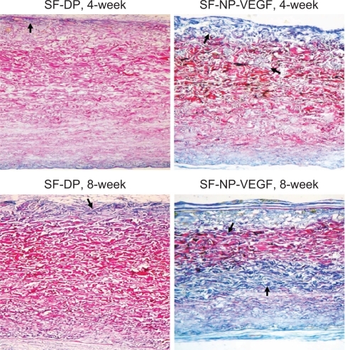
Figure 10 Glycosaminoglycans (GAG) staining. Scott’s alcian blue staining showed that GAGs were present in all layers of the wall, original magnification × 100. More blue staining (new collagen fibrils) was observed in SF-NP-VEGF implant at 4 and 8 week.
Note: The arrows indicate positive staining of new collagen fibrils.
Abbreviations: DP, decellularized scaffolds; NP, nanoparticle; SF, scaffolds; VEGF, vascular endothelial growth factor