Figures & data
Figure 1 Biocompatibility of G1-S4, G2-S16, and G1-S4/PD and G2-S16/PD combinations in TZM-bl epithelial cell line.
Notes: The viability of the TZM-bl cell line was evaluated by MTT assay 48 hours after exposure to these compounds, (A) PD, G1-S4, and G2-S16 alone, and (B) their combinations. Eighty percent of cell viability was established as a limit of toxicity, and 20 µM dextran and 10% DMSO were used as negative and positive cell death control, respectively. The data were represented as a mean ± standard deviation of four different experiments.
Abbreviations: NT, nontreated; PD, Platycodin D.
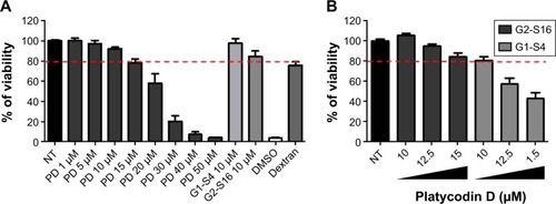
Figure 2 Dendrimers and combinations anti-HIV-1 activity.
Notes: (A) Inhibition of HIV-1 infection in TZM-bl. The TZM-bl were pretreated for 1 hour with nontoxic concentrations of the compounds and subsequently infected with HIV-1. The percentage of infection was determined after 48 hours by quantification of luciferase expression levels. (B) Inhibition of HIV-1 infection in PBMC. The PBMCs were pretreated for 1 hour with nontoxic concentrations of the compounds and subsequently infected with HIV-1. After 72 hours, the supernatants collected from PBMCs were presented on TZM-bl cells. The percentage of infection was determined after 48 hours by quantification of luciferase expression levels. Data were plotted as mean ± standard deviation of five different experiments. **P≤0.01; ***P≤0.001.
Abbreviations: NT, nontreated; PBMC, peripheral blood mononuclear cell; PD, Platycodin D.
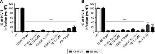
Figure 3 Time of addition assay.
Notes: To determine the stage within the HIV-1 viral cycle where PD and its combination with G1-S4 and G2-S16 carry out their inhibitory effect, TZM-bl cells were infected with HIV-1 and compounds were added at 0, 1, 2, 4, 6, and 8 hours postinfection. MVC, T-20, TDF, and RALT were used as controls of the HIV-1 infection. The percentage of viral infection was determined at 48 hours by quantification of luciferase expression levels. Data were plotted as mean ± standard deviation of three different experiments.
Abbreviations: MVC, maraviroc; NT, nontreated; PD, Platycodin D; RALT, raltegravir; T-20, enfuvirtide; TDF, tenofovir disopropil fumarate.
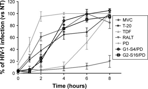
Figure 4 Cell proliferation assay in PBMC.
Notes: The ability of G1-S4 or G2-S16 dendrimers to PD to induce cell proliferation in PBMC was evaluated. PBMCs were treated with the compounds for 72 hours. Phytohemagglutinin 2 µg/mL was used as a positive proliferative control. The percentage of cell proliferation was determined using a Millipore proliferation kit. Data were plotted as mean ± standard deviation of three different experiments.
Abbreviations: NT, nontreated; PBMC, peripheral blood mononuclear cells; PD, Platycodin D.
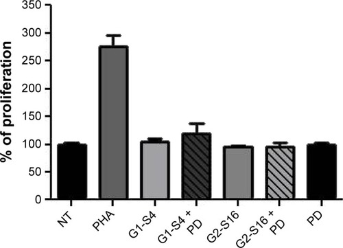
Figure 5 Sperm capacitation assay.
Notes: Human sperm samples, after being processed and selected by density gradient, were treated with G1-S4 or G2-S16 dendrimer in combination with PD. The mobile sperm count was evaluated to determine if the combination of the compounds tested affects sperm motility. The sperm motility index, which exceeds 70%, was considered optimal. Sperm from a single donor was used to perform the experiments.
Abbreviation: PD, Platycodin D.
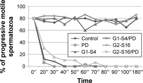
Table 1 Vaginal toxicity assay of G1-S16/PD or G2-16/PD after seven consecutive daily applications doses
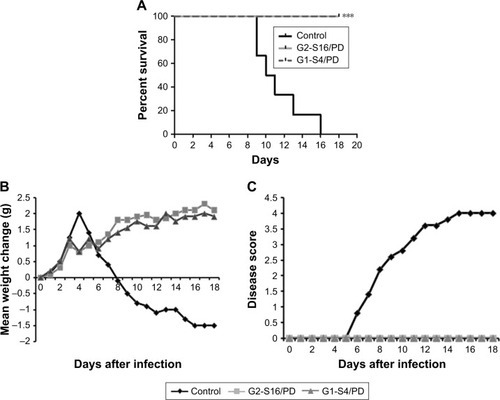
Figure 6 G1-S4 and G2-S16 dendrimers prevent vaginal high-dose HSV-2 infection in the presence of PD.
Notes: Medroxyprogesterone acetate-treated BALB/c mice were vaginally challenged with 105 PFU HSV-2 1 hour after applying the indicated gel (six mice/group). Mice were examined daily for body weight and genital pathology over 18 days. (A) Percentages of infection over time are shown for each treatment group. Dendrimer-based gels containing 3% dendrimer and 0.25 mM PD were significantly more protective than vehicle alone (***P<0.001 vs placebo). (B) Body weight changes were expressed as the mean values of ten animals in the same group. Each mean value was calculated by subtracting the weight at day 0 from the weight at day N after infection. (C) Clinical pathology was scored as described in the text for 18 days. Lesion scores were expressed as the mean values of six mice in the same group.
Abbreviations: HSV-2, herpes simplex virus type-2; PD, Platycodin D; PFU, plaque forming unit.