
Figures & data
Figure 1 The XRD pattern of ZVFe NP (A); FESEM image of ZVFe NPs (B); DLS histogram of ZVFe NPs (C).
Abbreviations: DLS, dynamic light scattering; FESEM, field-emission scanning electron microscope; XRD, X-ray diffraction; ZVFe NPs, zero valent iron nanoparticles.
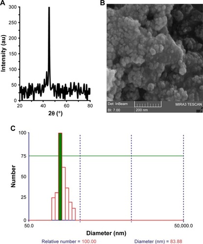
Figure 2 Fluorescence quenching of HSA upon interaction with ZVFe NP at three different temperatures: 298 K (A), 310 K (B), and 315 K (C).
Note: The fluorescence spectra of HSA (2 µM) in the presence of varying concentrations (0 [dark blue], 1 [brown], 2 [green], 4 [bright blue], 6 [purple], 8 [gray], 10 [bright brown], and 15 [red] µM) of ZVFe NPs.
Abbreviations: HSA, human serum albumin; ZVFe NPs, zero valent iron nanoparticles.
![Figure 2 Fluorescence quenching of HSA upon interaction with ZVFe NP at three different temperatures: 298 K (A), 310 K (B), and 315 K (C).Note: The fluorescence spectra of HSA (2 µM) in the presence of varying concentrations (0 [dark blue], 1 [brown], 2 [green], 4 [bright blue], 6 [purple], 8 [gray], 10 [bright brown], and 15 [red] µM) of ZVFe NPs.Abbreviations: HSA, human serum albumin; ZVFe NPs, zero valent iron nanoparticles.](/cms/asset/e960dcd0-65f6-4089-9851-d24d5747c274/dijn_a_12190680_f0002_c.jpg)
Table 1 KSV parameters at three different temperatures for the interaction of HSA with ZVFe NPs
Figure 3 Stern–Volmer plot of the fluorescence quenching of HSA (2 µM) in the presence of varying concentrations (1, 2, 4, 6, 8, 10, and 15 µM) of ZVFe NPs at 298 K (■), 310 K (♦), and 315 K (▲).
Abbreviations: HSA, human serum albumin; ZVFe NPs, zero valent iron nanoparticles.
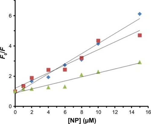
Table 2 Binding parameters at three different temperatures for the interaction of HSA with ZVFe NPs
Figure 4 Hill plot for the binding of HSA with ZVFe NPs at 298 K (■), 310 K (♦), and 315 K (▲).
Abbreviations: HSA, human serum albumin; ZVFe NPs, zero valent iron nanoparticles.
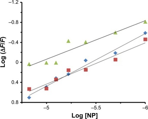
Table 3 Thermodynamic parameters at three different temperatures for the interaction of HSA with ZVFe NPs
Figure 5 Van’t Hoff plot for the binding of HSA with ZVFe NPs.
Abbreviations: HSA, human serum albumin; ZVFe NPs, zero valent iron nanoparticles.
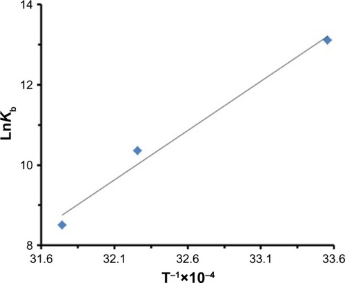
Figure 6 Far CD spectra of HSA (3 µM) in the presence of varying concentrations of ZVFe NP (0 [black], 3 [red], 10 [blue], and 30 [green] µM).
Abbreviations: CD, circular dichroism; HSA, human serum albumin; ZVFe NPs, zero valent iron nanoparticles.
![Figure 6 Far CD spectra of HSA (3 µM) in the presence of varying concentrations of ZVFe NP (0 [black], 3 [red], 10 [blue], and 30 [green] µM).Abbreviations: CD, circular dichroism; HSA, human serum albumin; ZVFe NPs, zero valent iron nanoparticles.](/cms/asset/6363c659-7635-4fc7-994c-193f3c508ab0/dijn_a_12190680_f0006_c.jpg)
Figure 7 The absorbance (295 nm) of HSA (3 µM) in the absence (blue line) and presence (red line) of ZVFe NP (3 M) over the temperature range of 30°C–90°C.
Abbreviations: HSA, human serum albumin; ZVFe NPs, zero valent iron nanoparticles.
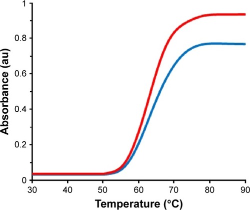
Figure 8 Molecular docking of HSA with water-coated ZVFe NP.
Abbreviations: HSA, human serum albumin; ZVFe NPs, zero valent iron nanoparticles.
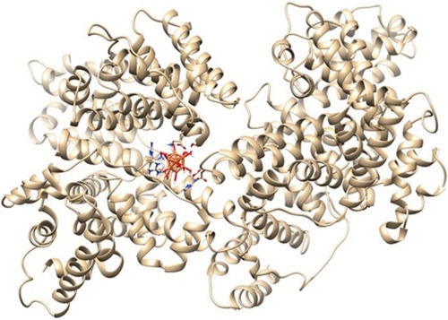
Figure 9 Different views (A–C) of the ligand and its surrounding site residues.
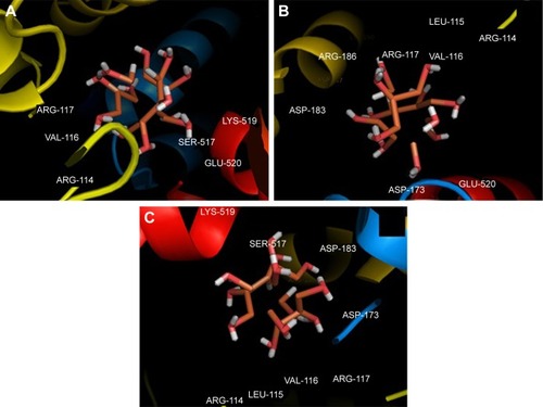
Figure 10 The viability percent of SH-SY5Y (blue bars) cells and WBCs (red bars) in the presence of varying concentrations of ZVFe NPs after 24 hours.
Note: *P<0.05 and **P<0.01 represent the significant differences between ZVFe NPs-treated groups and control.
Abbreviations: WBCs, white blood cells; ZVFe NPs, zero valent iron nanoparticles.
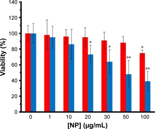
Figure 11 Flow cytometry analysis of ZVFe NP-induced apoptosis.
Notes: The induction of apoptosis was observed by IC50 concentration of ZVFe NPs (47.89±81 µg/mL) on SH-SY5Y cell. (A) Control cells and (B) treated group.
Abbreviation: ZVFe NPs, zero valent iron nanoparticles.
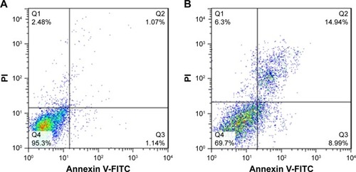
Table 4 MIC and MBC of ZVFe NP against three strains of bacteria
Figure 12 (A–C) Photographs of the zone of inhibition produced by ZVFe NP against three strains of bacteria.
Note: (A) Escherichia coli, (B) Pseudomonas aeruginosa, and (C) Staphylococcus aureus.
Abbreviation: ZVFe NPs, zero valent iron nanoparticles.
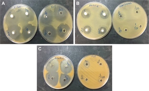
Figure 13 The antibacterial activity of ZVFe NPs aganist E.coli, P. aeruginosa and S. aureus bacterial strains tested by disc diffusion method. (Measuring inhibition zone diameter [mm]).
![Figure 13 The antibacterial activity of ZVFe NPs aganist E.coli, P. aeruginosa and S. aureus bacterial strains tested by disc diffusion method. (Measuring inhibition zone diameter [mm]).](/cms/asset/23b1a144-20af-4775-a1e2-313eaf4f0c21/dijn_a_12190680_f0013_c.jpg)