Figures & data
Figure 1 SEM image of the HA obtained.
Abbreviations: SEM, scanning electron microscopy; HA, hydroxyapatite.
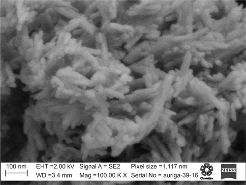
Figure 2 Chemical structure of APNPs.
Abbreviation: APNP, amphiphilic peptide nanoparticle.
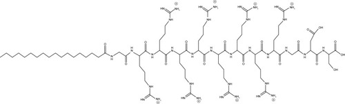
Figure 3 XRD pattern of the synthesized HA.
Abbreviations: XRD, X-ray diffraction; HA, hydroxyapatite.
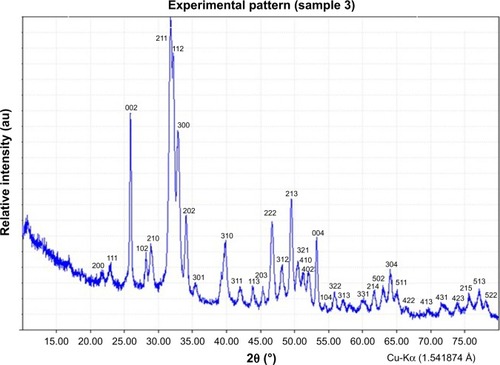
Figure 4 IR spectrum of the synthesized HA.
Abbreviations: IR, infrared; HA, hydroxyapatite.
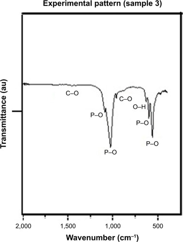
Figure 5 TEM images of (A–C) synthesized HA. (D) Electron diffraction pattern of HA.
Abbreviations: TEM, transmission electron microscope; HA, hydroxyapatite.
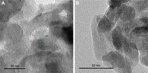
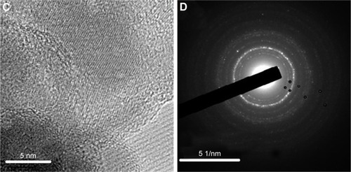
Table 1 Indexation of electron diffraction pattern
Figure 6 SEM images of (A) pHEMA coatings; (B) pHEMA + HA coatings; (C) pHEMA + APNPs coatings; and (D) pHEMA + HA + APNPs coatings.
Abbreviations: SEM, scanning electron microscope; pHEMA, 2-hydroxyethyl methacrylate; HA, hydroxyapatite; APNP, amphiphilic peptide nanoparticle.
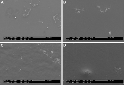
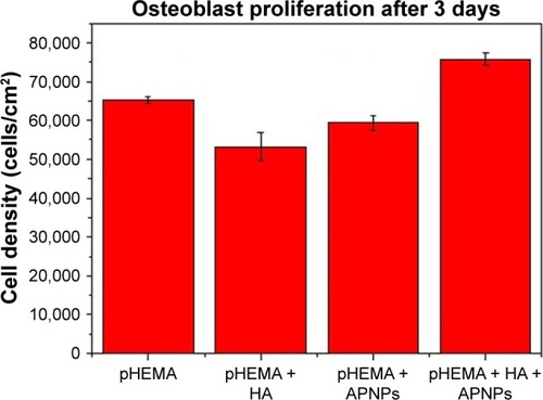
Figure 7 Osteoblast density on pHEMA, APNPs, and nanocrystalline HA-coated titanium. Data are mean ± S.E.M.; n=3. P<0.005 for all comparisons.
Abbreviations: pHEMA, 2-hydroxyethyl methacrylate; APNP, amphiphilic peptide nanoparticle; HA, hydroxyapatite; S.E.M., standard error of the mean.