Figures & data
Figure 1 (A) MDSC expression in S. aureus biofilm exposure. The bar graph (left panel) was illustrated and converted based on the flow cytometry results (right panel). (B) CURN suppressed biofilm-induced MDSC expansion in a dose-dependent manner. (C) CUR dissolved in PBS did not affect MDSC expansion. Data are expressed as the mean ± SD, *P<0.05 compared to biofilm-untreated group in (A). *P<0.05 and **P<0.01 compared to the biofilm-induced but no CURN treated group.
Abbreviations: CUR, curcumin; CURN, CUR nanoparticles; MDSCs, myeloid-derived suppressor cells; S. aureus, Staphylococcus aureus.
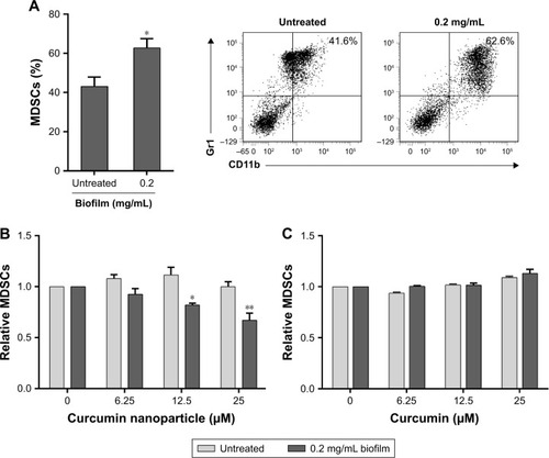
Figure 2 CURN induced expansion of MDSCs to M1- (A) and M2-phenotypic (B) MDSCs upon exposure to S. aureus biofilm via its anti-inflammatory activity.
Notes: Data are expressed as the mean ± SD. *P<0.05 and **P<0.01 compared to the biofilm-induced but no CURN treated group.
Abbreviations: CURN, curcumin nanoparticles; MDSCs, myeloid-derived suppressor cells; S. aureus, Staphylococcus aureus.
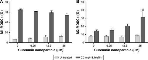
Figure 3 Cytokines including IL-6 (A), TNF-α (B), and MCP-1 (C) were downregulated by CURN in the mouse BMC culture supernatants of S. aureus biofilm-stimulated MDSCs.
Notes: Data are expressed as the mean ± SD. *P<0.05 and **P<0.01 compared to the biofilm-induced but no CURN treated group.
Abbreviations: BMCs, bone marrow cells; CURN, curcumin nanoparticles; MCP-1, monocyte chemoattractant protein-1; MDSCs, myeloid-derived suppressor cells; TNF-α, tumour necrosis factor-alpha; S. aureus, Staphylococcus aureus.
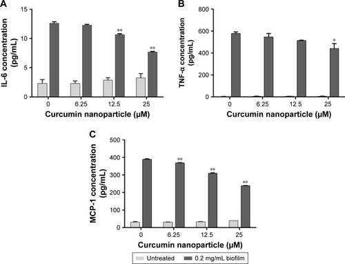
Figure 4 ROS generation from biofilm induced in mouse BMCs was suppressed by CURN in vitro.
Notes: DCF fluorescence was measured by flow cytometry. The bar graphs illustrate the signals of DCF fluorescence. Data are expressed as the mean ± SD. **P<0.01 compared to the biofilm-induced but no CURN treated group.
Abbreviations: BMCs, bone marrow cells; CURN, curcumin nanoparticles; DCF, dichlorofluorescin; ROS, reactive oxygen species.
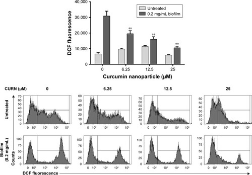
Figure 5 CURN reversed the immunosuppressive activity of S. aureus biofilm-treated MDSCs on T cells.
Notes: The differentiation rate of naive T cells after receiving CD3/CD28 beads stimulation was set as 100% (the mosaic bar). The changes in T-cell differentiation rate were measured after coculturing with MDSCs, which were pretreated with different concentrations of CURN and biofilm. Data are expressed as the mean ± SD. *P<0.05 and **P<0.01 compared to the biofilm-induced but no CURN treated group.
Abbreviations: CURN, curcumin nanoparticles; MDSCs, myeloid-derived suppressor cells; S. aureus, Staphylococcus aureus.
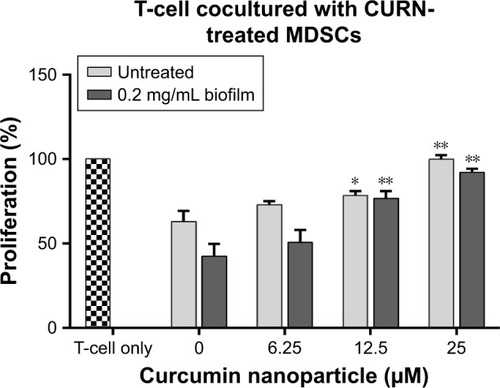
Figure 6 Evaluation of infection level in mouse femur.
Notes: (A) Micro-CT images, in which black arrows indicate the grade of severity in bone destruction: (i) sham group, (ii) CURN only, (iii) permeative destruction with S. aureus implantation, (iv) pathologic fracture after vancomycin (2 mg/kg) treatment, and (v–vii) the less osteolytic size in the gradient concentration of CURN. (B) Histological analysis by H&E staining showed that CURN was effective for treating PJIs cotreated with vancomycin (2 mg/kg). The labeling of figures is the same as that of micro-CT images. Red arrows indicate infiltrating mononuclear immune cells. (C) The inflammatory index score was evaluated based on the levels of inflammatory cell infiltration in the femur (n=3 for each group). Red arrows indicate infiltrating mononuclear immune cells. Data are expressed as the mean ± SD. *P<0.05 and **P<0.01 compared to the group of S. aureus infection treated with vancomycin alone.
Abbreviations: CT, computed tomography; CURN, curcumin nanoparticles; PJI, periprosthetic joint infection; S. aureus, Staphylococcus aureus.
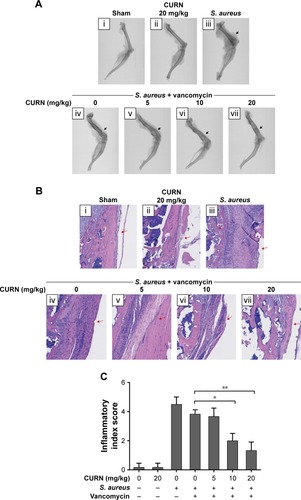
Figure 7 CURN reduces the expansion of MDSCs in the peripheral blood from mice infected with S. aureus.
Notes: Data are expressed as the mean ± SD. *P<0.05 compared to the group of S. aureus infection treated with vancomycin (2 mg/kg) alone.
Abbreviations: CURN, curcumin nanoparticles; MDSCs, myeloid-derived suppressor cells; S. aureus, Staphylococcus aureus.
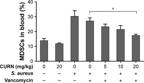
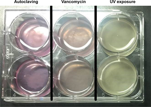
Figure S1 The supplementary data demonstrated that the autoclaving method for biofilm is the optimal method in our system.
Notes: S. aureus and its biofilm were collected by centrifugation at 3,900× g for 15 minutes, and then the pellet was sterilized by autoclaving or pre-treatment with vancomycin or UV for 12 hours (from lane 1 to lane 3) before adding to BMC culture. Both pre-treatment with vancomycin (lane 2) and UV for 12 hours (lane 3) failed to reduce the S. aureus growth rate. If the collected pellet was not autoclaved, the growth of S. aureus was rapid and resulted in the death of BMCs within 48 hours.
Abbreviations: BMCs, bone marrow cells; S. aureus, Staphylococcus aureus.