Figures & data
Figure 1 Osteochondral damage during the progression of OA.
Notes: OA subchondral bone is known to be hypomineralized and to show abnormal bone metabolism, resulting in histopathological degeneration of subchondral bone as well as cartilage damage. During progression of OA, the subchondral bone is also damaged as well as the articular cartilage layer. Injuries of either tissue adversely affect the mechanical environment as well as the homeostatic balance of the entire joint, resulting in the development of OA.
Abbreviation: OA, osteoarthritis.
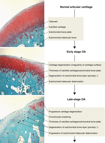
Table 1 International Cartilage Repair Society II scoring system for cartilage repair
Figure 2 Self-organized cartilage-like scaffold formed from HA, AG and type II collagen for articular cartilage tissue engineering.
Notes: (A) Macroscopic image of self-organized cartilage-like scaffold. (B) The structure of the self-organized AG/HA/collagen complex was observed by SEM. The topological structure of the complex had a fibrous appearance. (C) After 1- and 2-week incubation of chondrocytes with the self-organized AG/HA/collagen complex, chondrocytes were present on the scaffold of fibers forming the complex.
Abbreviations: AG, aggrecan; HA, hyaluronic acid; SEM, scanning electron microscopy.
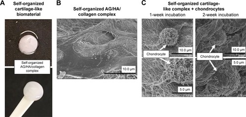
Figure 3 Self-organized biphasic cartilage and bone-like scaffold combined with an HAP bone block.
Notes: (A) Macroscopic image of our self-organized cartilage-like scaffold combined with an HAP block. (B) The structure of the self-organized cartilage-like scaffold combined with an HAP bone block was observed by SEM.
Abbreviations: HAP, hydroxyapatite; SEM, scanning electron microscopy.
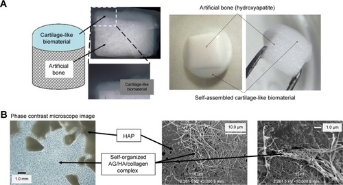
Figure 4 Self-assembled artificial cartilage–HAP powder conjugate.
Notes: (A) Diagram illustrating the formation of a self-assembled artificial cartilage–HAP powder conjugate and its macroscopic image. (B) The structure of the 40-nm size HAP powder coating complex was observed by SEM. (C) The structure of the 5-µm size HAP powder coating complex was observed by SEM.
Abbreviations: HAP, hydroxyapatite; SEM, scanning electron microscopy.
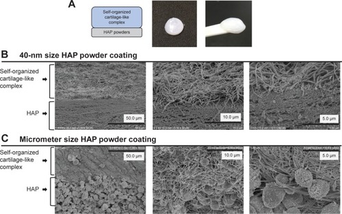
Figure 5 Viscoelasticity of self-organized biphasic cartilage and bone-like scaffold.
Notes: Only the artificial cartilage and the HAP-coated cartilage were measured and compared in terms of viscoelasticity by dividing it into three groups of wet, nondried and rehydrated. A significant difference between the two scaffolds was observed after rehydration. There were significant differences in the viscoelasticity between two groups as following: a vs c: P<0.01, b vs d: P<0.01, a vs e: P<0.01, and b vs f: P<0.01.
Abbreviation: HAP, hydroxyapatite.
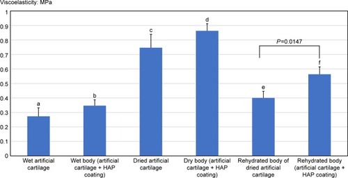
Figure 6 Safranin staining cross-sections of repaired articular cartilage and subchondral bone tissues in knee joins at 4 and 8 weeks after scaffold implantation.
Notes: (A) Representative images of control right knee joints and scaffold-implanted left knee joints at 4 weeks after scaffold implantation (control groups: n=3, each scaffold inoculated group: n=4). (B) Representative images of control right knee joints and scaffold-implanted left knee joints at 8 weeks after scaffold implantation (control groups: n=3, each scaffold-inoculated group: n=4).
Abbreviations: AG, aggrecan; HA, hyaluronic acid; HAP, hydroxyapatite.
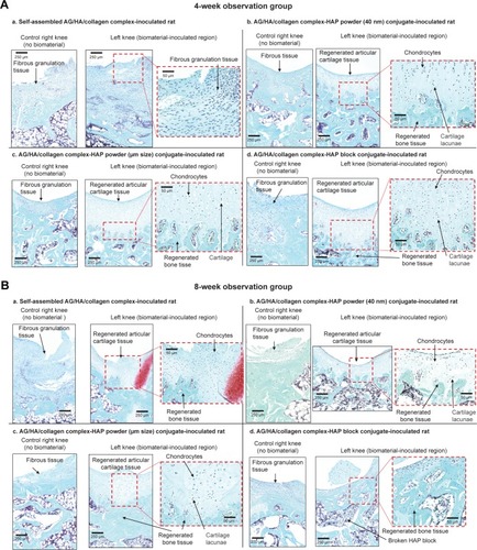
Figure 7 Histological assessment of osteochondral tissue regeneration by ICRS II scoring system.
Notes: (A) The histological quality of cartilage regeneration by scaffolds was evaluated by the ICRS II scoring system (). A higher score in the ICRS II scoring system represents tissue resembling healthy articular cartilage (control groups: n=3, each scaffold-inoculated group: n=4). (B) Histological assessment of subchondral bone regeneration was evaluated using parameters for subchondral abnormality in the ICRS II scoring system (control groups: n=3, each scaffold-inoculated group: n=4).
Abbreviations: HAP, hydroxyapatite; ICRS, International Cartilage Repair Society.

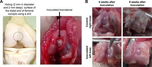
Figure 8 Implantation of self-organized scaffold for cartilage and subchondral bone tissue repair.
Notes: (A) Representative images: creation of the defect (2 mm in diameter and 2 mm deep) in the surface of the distal end of the femoral condyle using a drill. Implantation of the scaffold for osteochondral tissue repair. (B) Macroscopic images of the defect 4 and 8 weeks after implantation were compared between control and implanted groups.