Figures & data
Figure 1 Characterizations of BTO/P(VDF-TrFE) nanocomposite membranes. (A) Representative SEM image of the surface morphology of polarized nanocomposite membranes. (B) Cross-sectional SEM images of polarized composite membranes. Insets are the low magnification images. Yellow arrows denote the BTO nanoparticles. (C) X-Ray diffraction patterns of BTO/P(VDF-TrFE) nanocomposite membranes and neat P(VDF-TrFE) membranes. (D) Water contact angles of nanocomposite membrane before and after corona poling treatment. (E) The hysteresis loops of nanocomposite membranes. (F) The piezoelectric coefficient (d33) values of the polarized nanocomposite membranes after immersion in serum-free cell culture medium for different time durations.
Abbreviations: BTO, BaTiO3; P(VDF-TrFE), poly(vinylidene fluoridetrifluoroethylene); SEM, scanning electron microscopy.
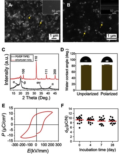
Figure 2 Osteogenic differentiation of BM-MSCs on polarized nanocomposite membranes and commercially-available PTFE membranes. (A) Representative immunostaining images for detection of RUNX-2 (green), actin network (red), and cell nuclei (DAPI, blue) in BM-MSCs cultured for 3 days. (B) Quantification of the immunostaining intensity of RUNX-2. Error bars represent one standard error. (**p<0.01).
Abbreviations: BM-MSCs, bone marrow mesenchymal stem cells; PTFE, polytetrafluoroethylene; RUNX-2, runt-related transcription factor 2; DAPI, 2-(4-amidinophenyl)-1H-indole-6-carboxamidine.
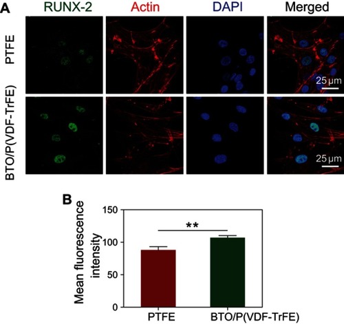
Figure 3 Illustration of the surgical procedure. (A) Bone defects with a diameter of 8-mm were created on rabbit mandibles, and then filled with DBB granules. (B) Bone defects were covered with polarized BTO/P(VDF-TrFE) nanocomposite membranes or commercially-available PTFE membranes.
Abbreviations: DBB, deproteinized bovine bone; BTO, BaTiO3; P(VDF-TrFE), poly(vinylidene fluoridetrifluoroethylene); PTFE, polytetrafluoroethylene.
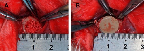
Figure 4 Histological evaluation of bone defects after implantation for 4 weeks and 12 weeks. (A, B, E, F) H&E staining images after 4 weeks of implantation. (A, E) PTFE membrane group. (B, F) BTO/P(VDF-TrFE) membrane group. (C, D, G, H) H&E staining images after 12 weeks of implantation. (C, G) PTFE membrane group. (D, H) BTO/P(VDF-TrFE) membrane group. The lower images are enlargements of specific regions of the upper images. (DBB: deproteinized bovine bone; NB: nascent bone; BM: bone marrow-like tissue). Scale bars =200 μm for (A–D); Scale bars =100 μm for (E-H). (I) Quantitative histomorphometry analysis of bone volume/total volume (BV/TV). Error bars represent one standard error. (**p<0.01).
Abbreviations: H&E, hematoxylin and eosin; PTFE, polytetrafluoroethylene; BTO, BaTiO3; P(VDF-TrFE), poly(vinylidene fluoridetrifluoroethylene); DBB, deproteinized bovine bone; NB, nascent bone; BM, bone marrow-like tissue.
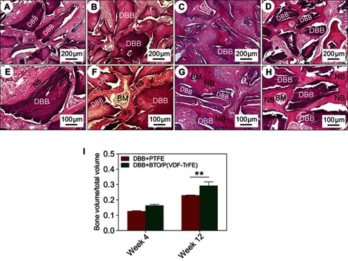
Figure 5 Histological observation of bone restoration after 12 weeks post-implantation. Representative Masson’s trichrome staining images of DBB granules within defects covered with PTFE membranes (A) or BTO/P(VDF-TrFE) nanocomposite membranes (B). Yellow arrows denote viable osteocytes in their lacunae. Scale bar =50 μm.
Abbreviations: DBB, deproteinized bovine bone; OT, osteoid tissue (red); MT, mineralized tissue (blue); PTFE, polytetrafluoroethylene; BTO, BaTiO3; P(VDF-TrFE), poly(vinylidene fluoridetrifluoroethylene).
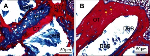
Figure 6 Evaluation of neovascularization at 4 weeks post-implantation. (A–B) Representative Masson’s trichrome staining images after implantation with DBB granules and covering with PTFE membranes (A) or BTO/P(VDF-TrFE) nanocomposite membranes (B). (C–D) Immunohistological staining images for detection of α-SMA expression after implantation with DBB granules and covering with PTFE membranes (C) or BTO/P(VDF-TrFE) nanocomposite membranes (D). (E) Quantitative analysis of the number of nascent blood vessels after 4 weeks post-implantation (**p<0.01). Black arrowheads denote the nascent blood vessels. Scale bar =50 μm.
Abbreviations: DBB, deproteinized bovine bone; NB, nascent bone; PTFE, polytetrafluoroethylene; BTO, BaTiO3; P(VDF-TrFE), poly(vinylidene fluoridetrifluoroethylene); α-SMA, alpha smooth muscle actin; HPF, high power fields.
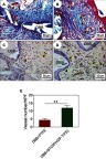
Figure 7 Immunohistochemical evaluation of bone regeneration at 12 weeks post-implantation. Representative immunohistochemical staining images for detection of OCN expression in DBB granules around nascent bone tissues covered with PTFE membranes (A) or BTO/P(VDF-TrFE) nanocomposite membranes (B). (C) and (D) are enlargements of specific regions of (A) and (B) respectively. Black arrowheads denote the positive expression of OCN. Scale bars =100 μm for (A–B); Scale bars =50 μm for (C-D).
Abbreviations: OCN, osteocalcin; DBB, deproteinized bovine bone; NB, nascent bone; PTFE, polytetrafluoroethylene; BTO, BaTiO3; P(VDF-TrFE), poly(vinylidene fluoridetrifluoroethylene).
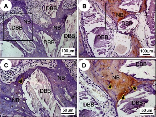
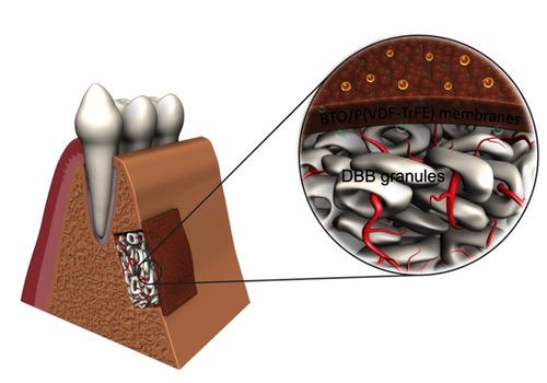
Figure 8 Illustration of the synergistic effects of combining piezoelectric BTO/P(VDF-TrFE) nanocomposite membranes with xenogenic DBB grafts on the repair of critical-sized bone defects. Electric dipoles of BTO NPs are reoriented in the direction of the poling electric field after corona poling treatment, and consequently polarized charges are generated on the surface of BTO/P(VDF-TrFE) nanocomposite membranes. When the membranes are implanted as a barrier membrane covering the bone defect filled with DBB grafts, the electric microenvironment is sustainably maintained, resulting in enhanced neovascularization and rapid bone regeneration, which consequently led to complete mature bone-structure formation integrated with the implanted DBB grafts.
Abbreviations: DBB, deproteinized bovine bone; BTO, BaTiO3; P(VDF-TrFE), poly(vinylidene fluoridetrifluoroethylene).