Figures & data
Figure 1 Schematic representation of different delivery systems. From left to right; liposomes, micelles, carbon nanotubes, dendrimer and gold (yellow) and iron (brown) nanoparticles.
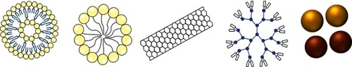
Figure 2 Image representing the blood transport mechanism of nanomaterials or molecules from normal tissue (left) and the enhanced permeability and retention effect in a tumor.
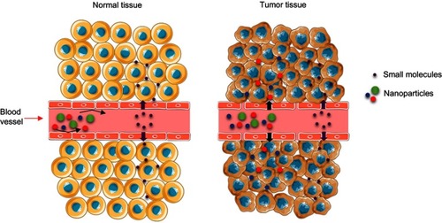
Figure 3 Schematic description of active targeting possibilities on mesoporous silica particles (left). Dual targeting example (right).
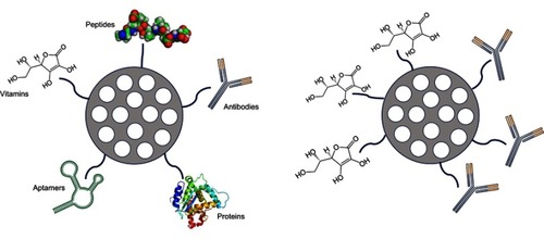
Table 1 Different gatekeepers that can be used to maintain the “zero release” of the drug and to trigger drug release
Figure 4 Examples of different gatekeepers that can be used to maintain the “zero release” of the drug inside mesoporous silica particles and to trigger on demand the release.
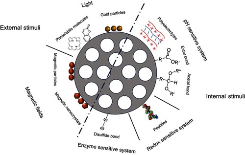
Figure 5 Scheme of the layer by layer technique in mesoporous silica particles.
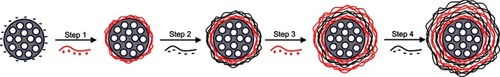
Figure 6 Diagram of the proton sponge effect: particles coated with polyethyleneimine (PEI) are captured in the endolysosomal route. Lysosomal membranes tear apart, releasing the particles in the cytosol.
Abbreviation: PEI-MSN, mesoporous silica particles coated with PEI.
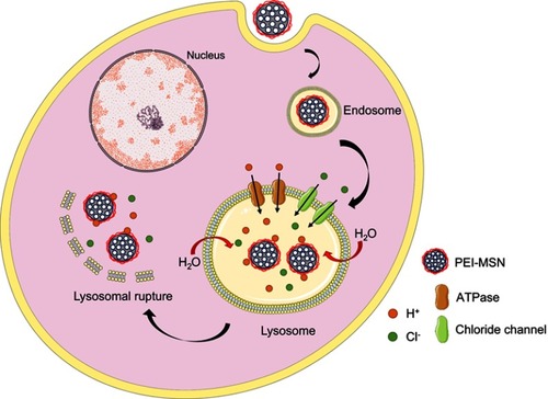
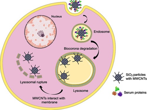
Figure 7 Diagram of how mesoporous SiO2 particles with a multi-walled carbon nanotubes (MWCNT) coating, scape the endolysosomal route. When proteins of the biocorona are degraded, apolar MWCNTs interact with the membrane and help particles escape these vesicles.