Figures & data
Figure 1 HRTEM images of green and orange CdTe nanoparticles showing an average size of 3 nm (A) and 5 nm (B), respectively.
Abbreviations: CdTe, cadmium telluride; HRTEM, high resolution transmission electron microscopy.
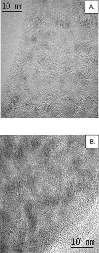
Figure 2 RSV-NP detection of RSV-infected Vero cells. Vero cells were mock-infected (A, C) or RSV-infected at a MOI = 1 (B, D) and fixed with acetone: methanol (60:40). Cells were immunostained with by conventional methods (A, B) or by RSV-NPs (C, D), and analyzed using an immunofluorescence microscopy (40X; A, C or 100X; B, D).
Abbreviations: MOI, multiplicity of infection; RSV-NP, respiratory syncytial virus-nanoparticles.
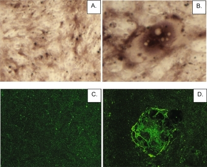
Figure 3 RSV-NP virus plaque assay. RSV was serially diluted ten-fold and the dilutions used to infect Vero cells for determination of virus titers. The viral titer determined from a conventional immunostaining plaque assay (A). were compared with RSV-NPs detection (B). Both 540 nm and 585 nm QDs were evaluated as RSV-NPs (B). RSV plaques were enumerated using an Amersham Biosciences Typhoon 9210 scanner.
Abbreviations: RSV-NP, respiratory syncytial virus-nanoparticles; QDs, quantum dots.
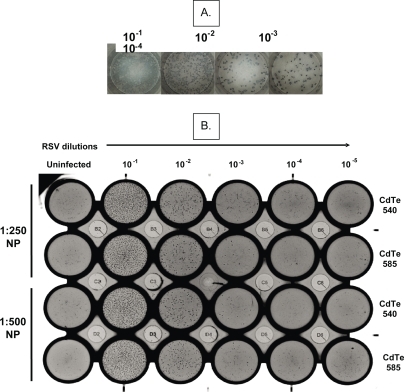
Figure 4 Dynamic range and sensitivity of detection of RSV-NP plaque assay compared with a conventional plaque assay. The viral titer of RSV-infected Vero cells was determined at day 3 pi (A) and day 5 pi (B) using conventional immunostaining plaque assay or single-step detection using 1:250 dilution of RSV-NPs derived from 585 nm QDs. RSV plaques were enumerated using an Amersham Biosciences Typhoon 9210 scanner.
Abbreviations: RSV-NP, respiratory syncytial virus-nanoparticles; QDs, quantum dots.
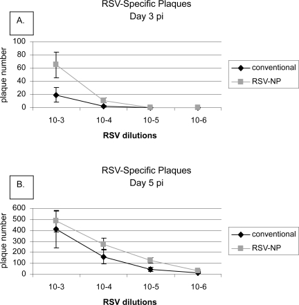
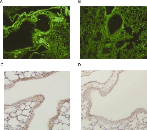
Figure 5 RSV-NPs detection of RSV-infected lung tissue. Lung tissue sections from RSV-infected (A, C) or naïve, mock-treated BALB/c mice (B, D) were stained by IHC using RSV-NPs (A, B), or by conventional IHC (C, D), and analyzed using an immunofluorescence microscopy.
Abbreviations: IHC, immunohistochemistry; RSV-NP, respiratory syncytial virus-nanoparticles.