Figures & data
Figure 1 NiNPs shape and size: (A) the shape of NiNPs as revealed by transmission electron micrographs of NiNPs. (B) Size distribution by number using a large number of TEM photos.
Abbreviations: NiNPs, nickel nanoparticles; SD, standard deviation; TEM, transmission electron microscope.
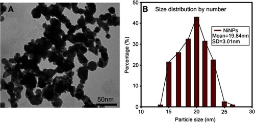
Figure 2 Internalization of NiNPs in rat kidney. (A) PT cell with different NiNPs localizations, within lysosome-like vacuoles (rectangle) and adhered to the basement membrane (circle and round curved rectangle) as shown in (B–D) respectively. Arrow indicates the lateral ridge of the basement membrane. (B) Lysosome-like vacuoles containing dense particles. (C) NiNPs (arrows) adhered to the basement membrane (BM). (D) NiNPs (arrows) either as individual particles inside the PT cell or adhered to the basement membrane of both adjacent PT cells. (E) Apical cytoplasmic region of PT cells showing discharged NiNPs (arrows) through the brush border into the lumen.
Abbreviations: PT, proximal convoluted tubule; NiNPs, nickel nanoparticles; BM, basement membrane; L, lysosome-like vacuoles; In, interstitial region; Ac, apical cytoplasmic region; BB, brush border; M, mitochondria; N, nucleus.
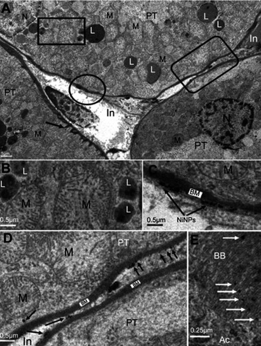
Table 1 Biochemical analysis in the serum of NiNP-treated rats
Figure 3 Internalization of NiNPs into the interstitial region of kidney cortex: (A) control group showing interstitial region between two proximal convoluted tubules. (B and C) the NiNP-treated group showing (B) deposition of NiNPs close to membranes (arrows), and (C) number of electron dense particles within vacuole close to the PT basement membrane (arrow).
Abbreviations: PT, proximal convoluted tubule; NiNPs, nickel nanoparticles; BM, basement membrane; In, interstitial region; M, mitochondria.
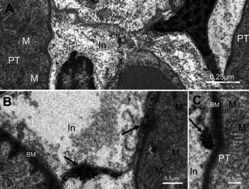
Figure 4 Basal region in proximal convoluted tubule cells of NiNP-treated rats. (A) Basal villi of control group (white arrow), lateral ridges (black arrows) extending from PT basement membrane (star). (B) Thickened basement membrane (star), disturbed basal villi (white arrows) and affected lateral ridges (black arrows).
Abbreviations: NiNPs, nickel nanoparticles; M, mitochondria; PT, proximal convoluted tubule.
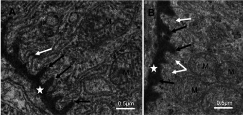
Figure 5 Ultrastructure of glomerulus after NiNPs administration: (A) control group showing well developed podocytes with their foot processes (arrow), endothelial and mesangial cells. Red blood cells in the blood capillaries are seen; (B) the NiNP-treated group showing degenerated podocytes and mesangial cells. The nuclei of mesangial cells (1) are seen fragmented.
Abbreviations: P, podocytes; EN, endothelial cells; RBC, red blood cells; BC, blood capillary; Mc, mesangial cells; NiNPs, nickel nanoparticles.
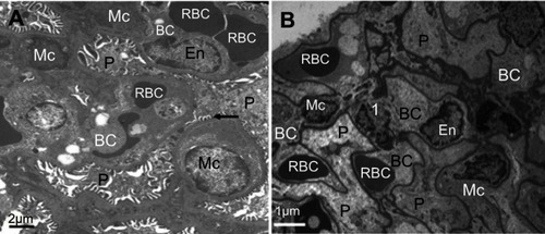
Figure 6 Ultrastructure changes in podocytes and mesangial cells in NiNP-treated rat kidney. (A) Degeneration of podocytes showing the fusion of foot processes (effacement) (arrows). Mesangial cells appeared degenerated having fragmented nucleus (black arrows). Neutrophil in the blood capillary showing phagocytosis of the degenerating mesangial cells. (B) Enlargement of (A) showing the phagocytosis process and engulfing of apoptotic bodies of mesangial cells and fusion of foot processes of podocytes.
Note: P1, primary podocyte foot processes; P2, secondary podocyte foot processes.
Abbreviations: NiNPs, nickel nanoparticles; P, podocytes; RBC, red blood cells; BC, blood capillary; MC, mesangial cells; Npl, neutrophil; ap, apoptotic bodies.
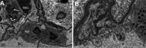
Figure 7 Immunohistochemical images of apoptotic cells in the glomerulus of NiNP-treated rat kidney. (A) Control group showing negative Tunel reaction. (B) NiNPs group showing positive Tunel reaction (arrows). (C) Control group showing no positive p53 reaction. (D) NiNPs group showing positive p53 reaction (arrows).
Abbreviations: NiNPs:, nickel nanoparticles; g, glomerulus.
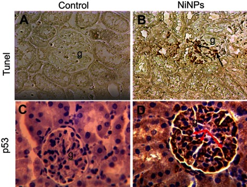
Figure 8 Ultrastructure changes in the podocytes foot processes. (A) Control group showing regular slit distances due to normal secondary foot processes structure (arrows; P2). (B) Fusion (effacement) of some of the secondary foot processes (P2). Arrow indicates a slit.
Note: P1, primary podocyte foot processes; P2, secondary podocyte foot processes.
Abbreviations: En, endothelial cells; RBC, red blood cells; BC, blood capillary; NiNPs, nickel nanoparticles; GBM, glomerular basement membrane.
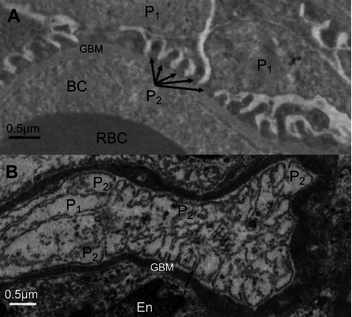
Figure 9 Ultrastructure of renal tubules of kidney of rats treated with NiNPs. (A) Proximal convoluted tubules of the NiNP-treated group showing variable sized lysosome-like vacuoles. Cellular debris is seen in the lumen of the duct shed from the renal tubules (stars). (B) Distal tubule of NiNP-treated rat showing pyknotic nuclei (arrows) against normal nuclei.
Abbreviations: NiNPs, nickel nanoparticles; L, lysosome-like vacuoles; N, nucleus; In, interstitial region.
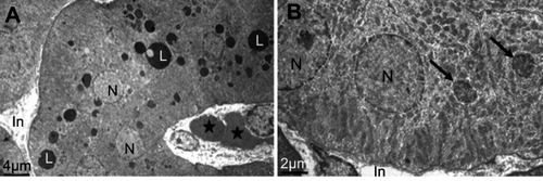
Figure 10 Electron microscope images of proximal convoluted tubule showing the status of mitochondria. (A) In the control group showing the normal density and ultrastructure. (B) In NiNP-treated rats showing the low density normal and damaged mitochondria. NiNPs are seen in different locations, free in the cytoplasm (black arrow), endocytosed by plasma membrane (white arrow), within lysosome-like vacuoles and uptaken by mitochondria (curved arrow connector).
Note: M2, damaged mitochondria.
Abbreviations: NiNPs, nickel nanoparticles; PT, proximal convoluted tubule; L, lysosome-like vacuoles; N, nucleus; M, mitochondria; bb, brush border.
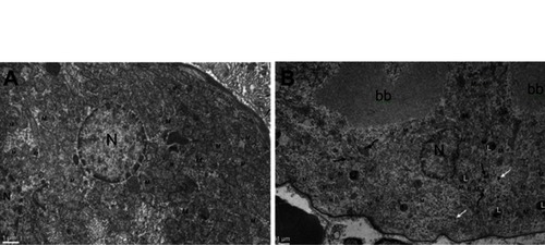
Figure 11 Mitochondria number counted by TEM photomicrographs per 100 µm2 area in PT cells of NiNPs treated rats which showed a significant decrease compared to control.
Note: *Significant decrease at P≤0.05.Abbreviation: NiNPs: nickel nanoparticles; TEM, transmission electron microscope; PT, proximal convoluted tubule.
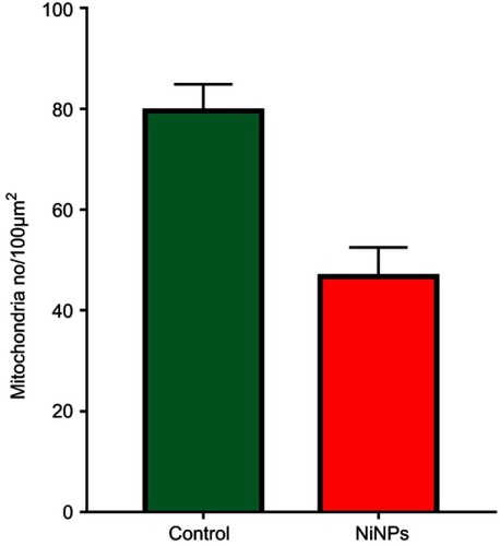
Figure 12 NiNPs intracellular localization in PT cells: (A) control group with normal ultrastructure of mitochondria, RER (arrow) and Golgi apparatus. (B) The NiNP-treated group showing electron dense particles, probably NiNPs, adhered to the RER which surround the mitochondria (arrows) and some are still free (arrow head).
Abbreviations: NiNPs, nickel nanoparticles; N, nucleus; RER, rough endoplasmic reticulum; M, mitochondria; PT, proximal convoluted tubule; G, Golgi apparatus.
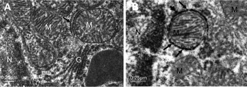
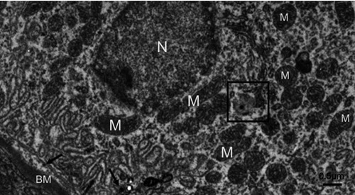
Figure 13 A distal tubule lining epithelial cell in the NiNP-treated group. Few numbers of mitochondria are seen. The box shows a vacuole containing an electron dense particle and whole vacuole is seen surrounded by an adjacent mitochondrion. Number of electron dense particles, probably NiNPs, near the basement membrane are seen (arrows).
Abbreviations: NiNPs, nickel nanoparticles; M, mitochondria; N, nucleus; BM, basement membrane.