Figures & data
Figure 1 Synthesis route of RGD-SWCNT(Rh)-thalidomide conjugates. (a) HNO3, 24 hours; (b) H2N-PEG-NH2, EDC, NHS, 0.1 M PBS, pH 7.4; (c) NHS-Rh, DMF; (d) SMCC, DMSO; (e) RGD-SH, thalidomide-SH.
Abbreviations: RGD, cyclic arginine-glycine-aspartic peptide; SWCNT, single-walled carbon nanotubes; Rh, rhodamine; PEG, polyethylene glycol; EDC, N-(3-dimethylaminopropyl)-3-ethylcarbodiimide hydrochloride; NHS, N-hydroxy succinimide; PBS, phosphate-buffered saline; DMF, dimethylformamide; SMCC, succinimidyl 4-[N-maleimidomethyl]cyclohexane-1-carboxylate; DMSO, dimethyl sulfoxide.
![Figure 1 Synthesis route of RGD-SWCNT(Rh)-thalidomide conjugates. (a) HNO3, 24 hours; (b) H2N-PEG-NH2, EDC, NHS, 0.1 M PBS, pH 7.4; (c) NHS-Rh, DMF; (d) SMCC, DMSO; (e) RGD-SH, thalidomide-SH.Abbreviations: RGD, cyclic arginine-glycine-aspartic peptide; SWCNT, single-walled carbon nanotubes; Rh, rhodamine; PEG, polyethylene glycol; EDC, N-(3-dimethylaminopropyl)-3-ethylcarbodiimide hydrochloride; NHS, N-hydroxy succinimide; PBS, phosphate-buffered saline; DMF, dimethylformamide; SMCC, succinimidyl 4-[N-maleimidomethyl]cyclohexane-1-carboxylate; DMSO, dimethyl sulfoxide.](/cms/asset/062e0f49-f3fd-46ed-b2a1-ba1027b02632/dijn_a_20145_f0001_b.jpg)
Figure 2 (A) Transmission electron microscopy images of pristine SWCNT; (B) thermogravimetric analysis curves of H2N-PEG-NH2 (dashed line), raw SWCNT (dotted line), and SWCNT-PEG-NH2 (solid line) under air which show the sequential loss of H2N-PEG-NH2 and SWCNT; (C) ultraviolet-visible absorbance spectra of PEGylated SWCNT, Rh, thalidomide, and RGD-SWCNT(Rh)-thalidomide conjugate 6 in H2O. The absorbance peak of thalidomide at 299 nm was used to measure the thalidomide loading on carbon nanotubes.
Abbreviations: RGD, cyclic arginine-glycine-aspartic peptide; SWCNT, single-walled carbon nanotubes; Rh, rhodamine; PEG, polyethylene glycol.
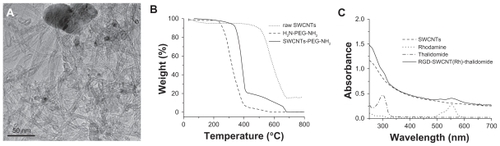
Figure 3 Confocal microscopic images of MCF7 cells (A, B and C) and U87MG cells (D, E and F) incubated with FITC-SWCNT (A and D) and FITC-SWCNT-RGD (B, C, E and F). (A) and (F) are the bright view images of (B) and (E), respectively. The green fluorescent signals indicate the location of FITC-SWCNT (A and D) and FITC-SWCNT-RGD (B and E) in the cells. Scale bar: 25 μm.
Abbreviations: RGD, cyclic arginine-glycine-aspartic peptide; SWCNT, single-walled carbon nanotubes; FITC, fluorescein isothiocyanate.
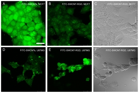
Figure 4 In vivo biodistribution of Rh-SWCNT-RGD in developing zebrafish embryos at different developmental stages. Zebrafish embryos were loaded with 2 nL of Rh-SWCNT-RGD (2.4 ng of SWCNT and 0.3 ng of RGD) into embryonic cells at the one-cell stage through microinjection. According to the red fluorescence signal from Rh-SWCNT-RGD, the loaded Rh-SWCNT-RGD are distributed into the blastoderm cells, but not the yolk cells, at all observed developmental stages, including the four-cell stage (A), 3 hours following fertilization (B), 8 hours following fertilization (C), and 28 hours following fertilization (D). Scale bar: 200 μm.
Abbreviations: RGD, cyclic arginine-glycine-aspartic peptide; SWCNT, single-walled carbon nanotubes; Rh, rhodamine.
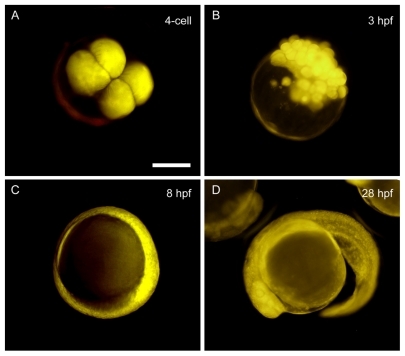
Figure 5 In vivo angiogenesis targeting of Rh-SWCNT-RGD in zebrafish wound healing model. By imaging transgenic fli1a:EGFP zebrafish that contained fluorescently-labeled endothelial cells, angiogenesis associated with wound healing in transgenic zebrafish embryos can be observed by checking the GFP signal. (A) Bright view and (B) Rh channel. Red fluorescence indicates the location of Rh-SWCNT-RGD in the zebrafish embryos. (C) indicates the FITC channel. Green fluorescence indicates distribution of fluorescently-labeled endothelial cells in transgenic zebrafish embryos. The red boxed area marks the region with a wound. The green boxed area marks a normal region in the zebrafish trunk. Scale bar: 150 μm.
Abbreviations: RGD, cyclic arginine-glycine-aspartic peptide; SWCNT, single-walled carbon nanotubes; Rh, rhodamine; FITC, fluorescein isothiocyanate; EGFP, enhanced green fluorescent protein.
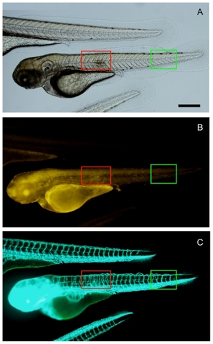
Figure 6 Inhibitory effects of SWCNT-conjugated thalidomide on angiogenesis in transgenic zebrafish embryos. Transgenic fli1a:EGFP zebrafish embryos were treated with 0.334 mg/mg thalidomide-SWCNT (thalidomide concentration, 1.2 mg/mL of SWCNT) from 4 hours following fertilization to 48 hours following fertilization, and then visualized live under a Leica SPE confocal microscope. The absence (A) and thinning (B) of intersegmental blood vessels, as indicated by the white arrows, is randomly distributed in zebrafish embryos after treatment with SWCNT-thalidomide. Scale bar: 250 μm in (A) and 100 μm in (B).
Abbreviation: SWCNT, single-walled carbon nanotubes.
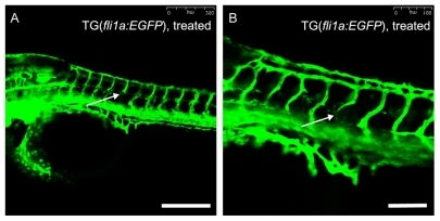
Figure 7 Tumor xenograft model in zebrafish embryos. Zebrafish embryos have been microinjected with fluorescent-labeled SKOV3:pEGFP mammalian cells suspended in Matrigel. The microinjection was conducted through the perivitelline space between the yolk and the periderm, close to the developing SIVs. The injection site is indicated by bright view in (A) using a white arrow, green fluorescence in (B) indicates presence of fluorescent-labeled SKOV3:pEGFP mammalian cells after injection, red fluorescence in (C) indicates presence of RGD-SWCNT(Rh)-thalidomide after injection, and (D) is (B) and (C) merged together. Scale bar: 150 μm.
Abbreviations: RGD, cyclic arginine-glycine-aspartic peptide; SWCNT, single-walled carbon nanotubes; Rh, rhodamine; EGFP, enhanced green fluorescent protein; SIVs, subintestinal vessels.
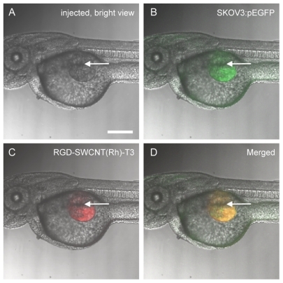
Figure 8 Blood vessels of transgenic fli1a:EGFP zebrafish embryos can be easily observed under the confocal microscope (A), and SIVs are marked by white arrows. Angiogenic responses (B and C) are triggered by tumor cell xenografts and targeted antiangiogenic therapy of RGD-SWCNT(Rh)-thalidomide (D, E and F) in transgenic fli1a:EGFP zebrafish embryos. Engraftment of human HT1080 fibrosarcoma cells, which secrete vascular endothelial growth factors, triggers ectopic angiogenesis of SIVs (B and C). Note morphological features of angiogenic response with engraftment of human HT1080 fibrosarcoma cells. When coinjected with RGD-SWCNT(Rh)-thalidomide (E), ectopic growth of angiogenesis of the SIV is obviously inhibited (D and F) in treated zebrafish embryos. White arrows (E and F) indicate presence of RGD-SWCNT(Rh)-thalidomide after injection. (F) is the merge of (D) and (E). Scale bar: 200 μm.
Abbreviations: RGD, cyclic arginine-glycine-aspartic peptide; SWCNT, single-walled carbon nanotubes; Rh, rhodamine; EGFP, enhanced green fluorescent protein; SIVs, subintestinal vessels.
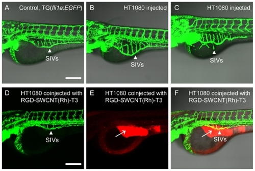
Figure 9 Total SIV length and area measurements in the zebrafish angiogenesis assay. The transgenic fli1a:EGFP zebrafish was used to study in vivo angiogenesis. By imaging live transgenic fli1a:EGFP zebrafish that contained fluorescently labeled endothelial cells at 72 hours following fertilization, the blood vessels are visualized under confocal microscopy. The total SIV vessel length and area is determined by using the NIH Image program. Each bar represents the mean ± standard deviation (n = 15). The control group refers to embryos injected with Matrigel only, the xenografted group refers to embryos xenografted with proangiogenic mammalian tumor cells HT1080, and the xenografted and treated group refers to embryos xenografted with proangiogenic tumor cells together with antiangiogenic nanotherapeutics RGD-SWCNT(Rh)-thalidomide.
Abbreviations: RGD, cyclic arginine-glycine-aspartic peptide; SWCNT, single-walled carbon nanotubes; Rh, rhodamine; EGFP, enhanced green fluorescent protein; SIV, subintestinal vessel.
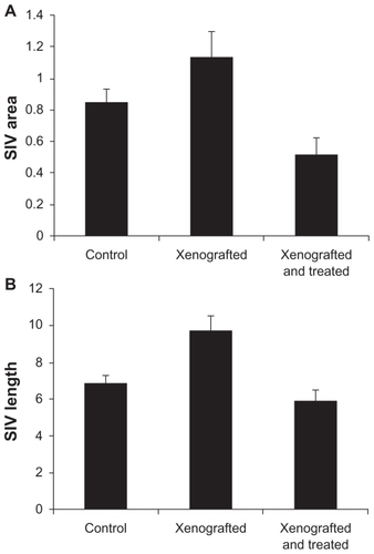
Figure S1 Synthesis of thalidomide analogs. Reagents and conditions are the following: (a) 2-(Boc-amino) ethyl bromide, trifluoroacetic acid, room temperature, 12 hours; (b) trifluoroacetic acid in CH2Cl2, 4 hours; (c) SATA, phosphate-buffered saline, 2 hours; (d) NH2OH, ethylenediamine tetra-acetic acid, phosphate-buffered saline.
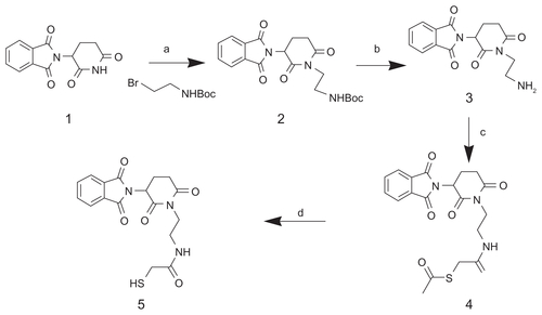
Figure S2 Synthesis route of SWCNT-FITC 7 and FITC-SWCNT-RGD 9 conjugates. (a) HNO3, 24 hours; (b) H2N-PEG-NH2, EDC, NHS, 0.1 M PBS, pH 7.4; (c) FITC, DMF, dark; (d) SMCC, DMSO; (e) RGD-SH, 0.1 M PBS.
Abbreviations: FITC, fluorescein isothiocyanate; RGD, cyclic arginine-glycine-aspartic peptide; SWCNT, single-walled carbon nanotubes; SMCC, succinimidyl 4-[Nmaleimidomethyl] cyclohexane-1-carboxylate; DMSO, dimethyl sulfoxide; PBS, phosphate-buffered solution; EDC, 1-ethyl-3-(3-dimethylamino-propyl) carbodiimide; PEG, polyethylene glycol; DMF, dimethylformamide.
![Figure S2 Synthesis route of SWCNT-FITC 7 and FITC-SWCNT-RGD 9 conjugates. (a) HNO3, 24 hours; (b) H2N-PEG-NH2, EDC, NHS, 0.1 M PBS, pH 7.4; (c) FITC, DMF, dark; (d) SMCC, DMSO; (e) RGD-SH, 0.1 M PBS.Abbreviations: FITC, fluorescein isothiocyanate; RGD, cyclic arginine-glycine-aspartic peptide; SWCNT, single-walled carbon nanotubes; SMCC, succinimidyl 4-[Nmaleimidomethyl] cyclohexane-1-carboxylate; DMSO, dimethyl sulfoxide; PBS, phosphate-buffered solution; EDC, 1-ethyl-3-(3-dimethylamino-propyl) carbodiimide; PEG, polyethylene glycol; DMF, dimethylformamide.](/cms/asset/d3e82f5a-1eac-4214-9f54-df6c631ceb51/dijn_a_20145_sf0002_b.jpg)