Figures & data
Table 1 BET analysis of CHA powder: pore diameter, cumulative pore volume, and cumulative surface area
Figure 1 (A) X-ray diffractograms and (B) infrared spectra of CHA microspheres and minocycline-loaded CHA microspheres.
Abbreviation: CHA, carbonated hydroxyapatite.
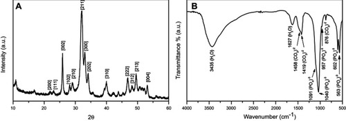
Figure 2 SEM micrographs of cross-section of (A) CHA and (B) CHAMINO microspheres, and (C) surface of CHA and (D) CHAMINO microspheres.
Notes: (A) and (B) magnification =250X (scale bar=400µm); (C) and (D) magnification=10,000X (scale bar=10µm).
Abbreviations: SEM, Scanning Electron Microscopy; CHA, Carbonated hydroxyapatite; CHAMINO, Minocycline-loaded nanocristalline carbonated hydroxyapatite.
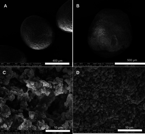
Figure 3 SR-µCT of CHA microsphere: (A) VR of microsphere with normalized orthoprojections; (B) orthoslice showing porous space inside the sphere; (C) VR of porous space inside sphere; (D) box representing individual pores of central microsphere region.
Abbreviations: orthoslice, orthogonal slice; SR-µCT, synchrotron radiation-based X ray microtomography; CHA, carbonated hydroxyapatite; VR, volume rendering.
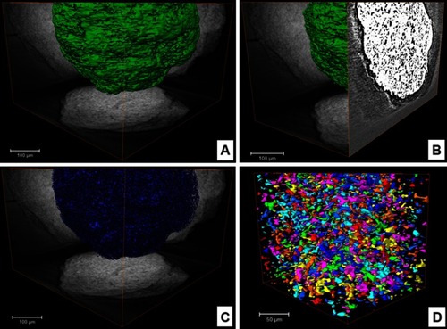
Figure 4 Cumulative MINO (%) release from CHA microspheres in PBS; the MIC values (mg/mL) of the microspheres at 1, 3, 5, and 7 days before and after the MINO release are shown for the E. faecalis culture.
Abbreviations: PBS, phosphate-buffered saline solution; MIC, minimum inhibitory concentrations.
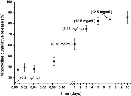
Figure 5 In vitro cell viability using F-OST cells cultured in extracts obtained from CHA and CHAMINO powders and microspheres. Cells seeded over Thermanox coverslip DMEM medium supplemented with 10% FBS were used as the negative control (C-), and 1% sodium dodecyl sulfate (SDS) and MINO 0.25% were used as the positive control, respectively (C+; MINO). Statistical analysis consisted of one-way ANOVA with Dunnett’s post hoc test (*p<0.001).
Abbreviations: DMEM, Dulbecco’s modified essential medium; FBS, fetal bovine serum; SDS, sodium dodecyl sulfate.
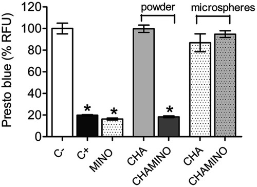
Figure 6 (A) One-week CHA and (B) CHAMINO groups. After 7 days of implantation, the presence of biomaterial microspheres was observed (B) surrounded by connective tissue (CT) and with newly formed bone in the CHAMINO group (*). Magnification: 40X; Stain: Hematoxylin and Eosin.
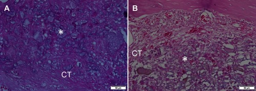
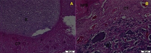
Figure 7 Six-week (A) of nanocrystalline carbonated hydroxyapatite (CHA) and (B) minocycline-loaded nanocrystalline carbonated hydroxyapatite (CHAMINO) groups. After 42 days of implantation, the residual particles of the biomaterial microspheres were observed surrounded by newly formed bone and connective tissue areas in both groups. Magnification: 40×; Stain: hematoxylin and eosin.
Abbreviations: CT, connective tissue; B, biomaterial; NFB, newly formed bone.
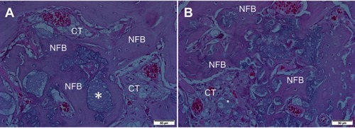
Figure 8 Histomorphometric evaluation of extraction sites following implantation of nanostructured carbonated hydroxyapatite microspheres. Connective tissue (green); biomaterial (yellow); newly formed bone (blue); other (orange).
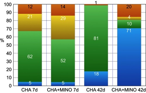
Figure S1 Equivalent pore diameter distribution of nanocrystalline carbonated hydroxyapatite (CHA) microsphere determined by SR-µCT.
Abbreviations: SR-µCT, synchrotron radiation-based X ray microtomography.
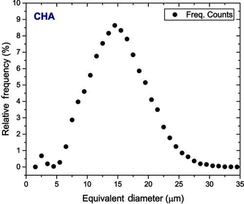
Figure S2 (A) One-week nanocrystalline carbonated hydroxyapatite (CHA) and (B) minocycline-loaded nanocristalline carbonated hydroxyapatite (CHAMINO) groups. After one week of implantation, the presence of non-degraded CHA microspheres was observed surrounded by connective tissue and with newly formed bone in the CHAMINO group. Magnification: 40×; stain: Hematoxylin and Eosin.
Abbreviations: B, biomaterial; NFB, newly formed bone; CT, connective tissue.