Figures & data
Table 1 Characterization of nanoparticles
Figure 1 Transmission electron microscopy images of 5-FU-entrapped PLGA 50-50 (A) and PLGA 90-10 nanoparticles (B).
Abbreviations: FU, 5-fluorouracil; PLGA, poly (D, L-lactic-co-glycolic acid).
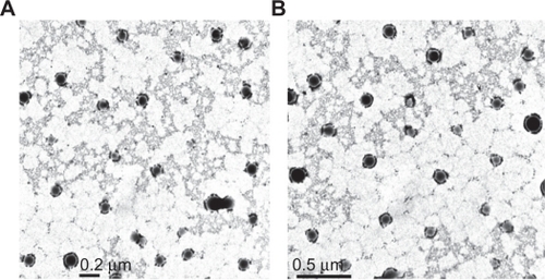
Figure 2 Polydispersity graphs of 5-FU-entrapped PLGA 50-50 (A) and PLGA 90-10 nanoparticles (B).
Abbreviations: FU, 5-fluorouracil; PLGA, poly (D, L-lactic-co-glycolic acid).
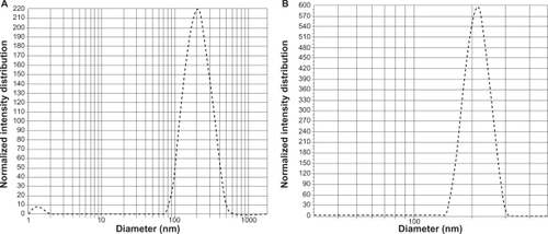
Figure 3 Differential scanning calorimetry thermograms of free 5-FU (A), vacant 5-FU PLGA nanoparticles (B), and 5-FU-entrapped PLGA nanoparticles (C).
Abbreviations: FU, 5-fluorouracil; PLGA, poly (D, L-lactic-co-glycolic acid).
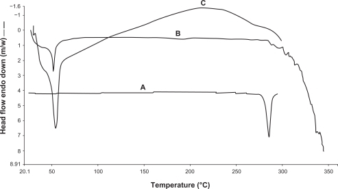
Figure 4 X-ray diffractograms of free 5-FU (A), vacant PLGA 50-50 nanoparticles (B), and 5-FU entrapped PLGA 50-50 nanoparticles (C).
Abbreviations: FU, 5-fluorouracil; PLGA, poly (D, L-lactic-co-glycolic acid).
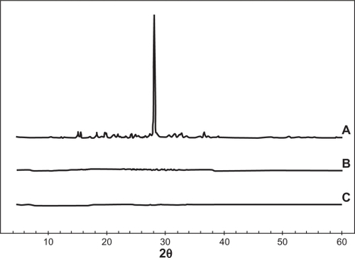
Figure 5 In-vitro drug-release pattern of 5-FU from 5-FU-entrapped PLGA nanoparticles.
Abbreviations: FU, 5-fluorouracil; PLGA, poly (D, L-lactic-co-glycolic acid).
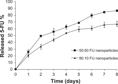
Figure 6 Confocal images of U87MG cells after 2 hours incubation with rhodamine-entrapped PLGA NPs.
Abbreviations: DAPI, 4′,6-diamidino-2-phenylindole; NP, nanoparticle; PLGA, poly (D, L-lactic-co-glycolic acid).
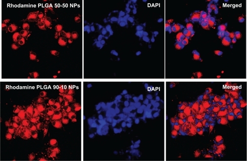
Figure 7 Confocal images of MCF7 cells after 2 hours incubation with rhodamine-entrapped PLGA NPs.
Abbreviations: DAPI, 4′,6-diamidino-2-phenylindole; NP, nanoparticle; PLGA, poly (D, L-lactic-co-glycolic acid).
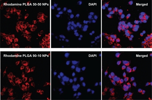
Figure 8 Cell viability of U87MG cells treated with 5-FU-entrapped PLGA 50-50 and 90-10 NPs compared with free 5-FU (mean ± standard deviation; n = 6).
Abbreviations: Conc, concentration; FU, 5-fluorouracil; NP, nanoparticle; PLGA, poly (D, L-lactic-co-glycolic acid).
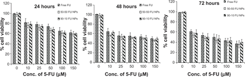
Figure 9 Cell viability of MCF7 cells treated with 5-FU-entrapped PLGA 50-50 and 90-10 NPs compared with free 5-FU (mean ± standard deviation; n = 6).
Abbreviations: Conc, concentration; FU, 5-fluorouracil; NP, nanoparticle; PLGA, poly (D, L-lactic-co-glycolic acid).
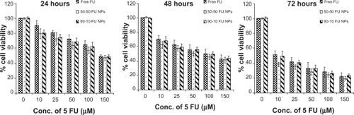
Figure 10 Fluorescence images of U87MG cells stained with acridine orange/ethidium bromide followed by quantification of apoptosis based on morphology changes.
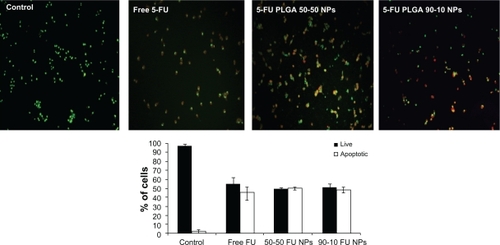
Figure 11 Fluorescence images of MCF7 cells stained with acridine orange/ethidium bromide followed by quantification of apoptosis based on morphology changes.
Abbreviations: FU, 5-fluorouracil; NP, nanoparticle; PLGA, poly (D, L-lactic-co-glycolic acid).
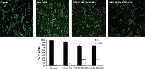
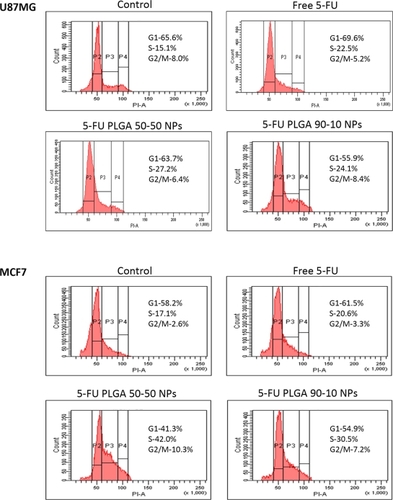
Figure 12 Cell-cycle analysis of U87MG (A) and MCF7 (B) cells on treatment with different 5-FU formulations.
Abbreviations: FU, 5-fluorouracil; NP, nanoparticle; PLGA, poly (D, L-lactic-co-glycolic acid).