Figures & data
Table 1 Composition of acetazolamide nanovesicles
Table 2 The physicochemical characterization of acetazolamide loaded nanovesicles
Figure 1 Transmission electron microscope (TEM) micrographs of prepared acetazolamide loaded nanovesicles. (A) TEM micrograph of F2 (Span 60:Tween 80 ratio 80:20). (B) TEM micrograph of F5 (Span 60:sodium deoxycholate ratio 80:20).
Abbreviations: F, formulation; Span 60, sorbitan monostearate.
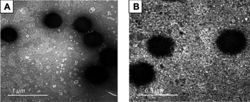
Table 3 Composition and characterization of ACZ-NV nanogels
Figure 2 In vitro release profile of acetazolamide from acetazolamide loaded nanovesicles (ACZ-NVs) and ACZ-NVs loaded in Chitosan nanogel.
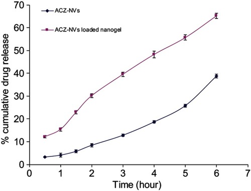
Table 4 The entrapment efficiency (EE) measurements (%) and particle size of the prepared nanovesicles during storage at 4°C, 25°C, and 37°C over a period of 3 months
Table 5 The percent drug content (%) of the prepared nanogel during storage at 4°C, 25°C, and 40°C/75%±5% relative humidity over a period of 1 month
Figure 3 Effect of different formulations of acetazolamide on lowering intraocular pressure.
Abbreviation: ACZ-NV, acetazolamide loaded nanovesicle.
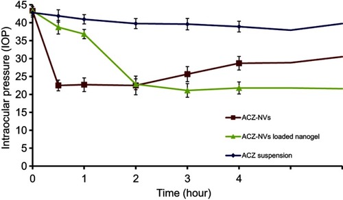
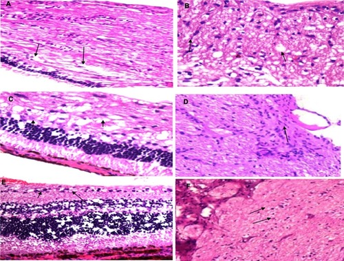
Figure 4 Histopathological examination; findings were appointed with the arrows shown in each image. (A) Glaucoma retina (retina showing loss of internal nuclear layer and ganglion cells with numerous numbers of vacuoles in the choroid). (B) Glaucomatous optic nerve (optic nerve showing thickening, demyelination, and accumulation of fat vacuoles). (C) Glaucoma retina treated with acetazolamide (ACZ) oral tablet (retina showing some loss of inner plexiform layer and ganglion cells with little vacuoles in choroid). (D) Glaucomatous optic nerve treated with ACZ oral tablet (optic nerve showing Wallerian degeneration and minimal loss of myelin sheath). (E) Glaucoma retina treated with ACZ nanovesicles loaded in Chitosan nanogel (retina showing mild swelling of ganglion cells). (F) Glaucomatous optic nerve treated with ACZ nanovesicles loaded in Chitosan nanogel (optic nerve showing mild edema and thickening with remyelination of nerve fibers). Magnification power was 100×.