Figures & data
Scheme 1 Schematic illustration of the preparation and application of MSN-DPH (consist of Dox loaded, TPP attached, HA capped mesoporous silica nanoparticles) for multistage-targeted delivery of Dox.
Abbreviations: Dox, Doxorubicin; HA, hyaluronic acid; MSN, mesoporous silica nanoparticle; TPP, triphenylphosphine.
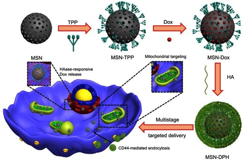
Figure 1 Characterizations of the MSNs and MSN-DPH (consist of Dox loaded, TPP attached, HA capped mesoporous silica nanoparticles). Transmission electron microscopy images of (A) MSN and (B) MSN-DPH. Hydrodynamic size distribution of MSN (C) and MSN-DPH (D).
Abbreviation: MSN, mesoporous silica nanoparticle.
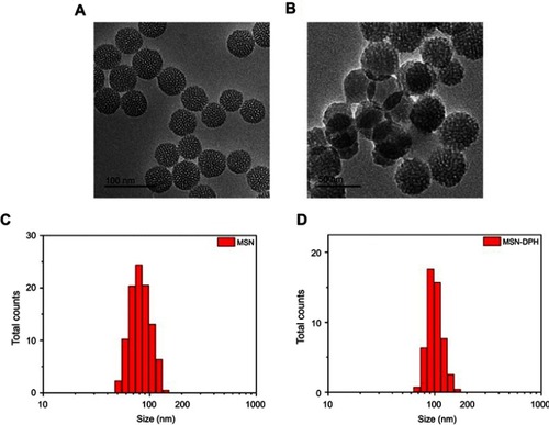
Figure 2 Characterizations of the MSN-DPH (consist of Dox loaded, TPP attached, HA capped mesoporous silica nanoparticles). (A) FT-IR spectra of MSN, MSN-NH2 and MSN-TPP. (B) Zeta potentials of MSN, MSN-NH2, MSN-TPP and MSN-DPH. Nitrogen adsorption–desorption isotherms (C) and corresponding pore size distribution (D) of MSN and MSN-DPH. (E) Stability of MSN-DPH in terms of hydrodynamic size in PBS and medium.
Abbreviations: MSN, mesoporous silica nanoparticle; TPP, triphenylphosphine.

Figure 3 Cumulative release of Dox from MSN-Dox, MSN-DPH (consist of Dox loaded, TPP attached, HA capped mesoporous silica nanoparticles) and MSN-DPH with the presence of HAase versus time. Data were presented as mean±SD (n=3).
Abbreviations: HAase, hyaluronidase; Dox, Doxorubicin; MSN, mesoporous silica nanoparticle.
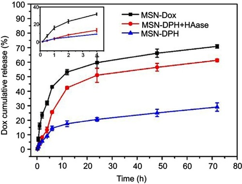
Figure 4 Cellular uptake and intracellular localization of MSN-DPH (consist of Dox loaded, TPP attached, HA capped mesoporous silica nanoparticles). Flow cytometry profiles of MGC-803 cells incubated with MSN-Dox (A) and MSN-DPH (B) for various time. (C) Confocal microscopy images of MGC-803 cells incubated with MSN-DPH for various time. The nuclei of the cells were stained with DAPI (blue). Cell mitochondria was stained by Mitolite Green. Scale bars denote 25 μm.
Abbreviations: Dox, Doxorubicin; MSN, mesoporous silica nanoparticle.
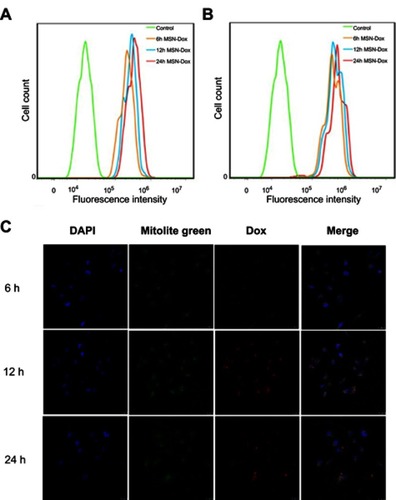
Figure 5 Relative viability of (A) Cos-7 and (B) MGC-803 cells incubated with MSN and MSN-DPH (consist of Dox loaded, TPP attached, HA capped mesoporous silica nanoparticles) for 48 hrs. Data were presented as mean±SD (n=3).
Abbreviation: MSN, mesoporous silica nanoparticle.
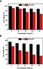
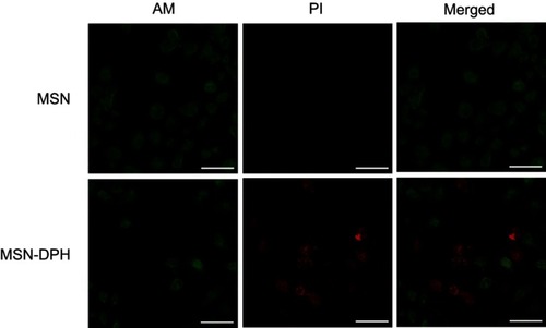
Figure 6 Live/dead images of MGC-803 cells incubated with MSN and MSN-DPH (consist of Dox loaded, TPP attached, HA capped mesoporous silica nanoparticles). Live cells and dead cells were stained by AM (green) and PI (red), respectively. Scale bars denote 25 μm.
Abbreviation: MSN, mesoporous silica nanoparticle.