Figures & data
Figure 1 (A) Schematic of the experimental setup to co-axially electrospin the sheath-core-structured nanofibers; (B) photo of the co-axial nozzle and needles used for the experiments.
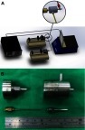
Figure 2 SEM image and fiber size distribution of lidocaine/hEGF-loaded nanofibers.
Abbreviations: SEM, scanning electron microscope; hEGF, human epidermal growth factor.
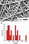
Figure 3 Laser scanning confocal microscopy images of reGFP in co-axial electrospun nanofibers.
Abbreviation: reGFP, recombinant enhanced green fluorescent protein.
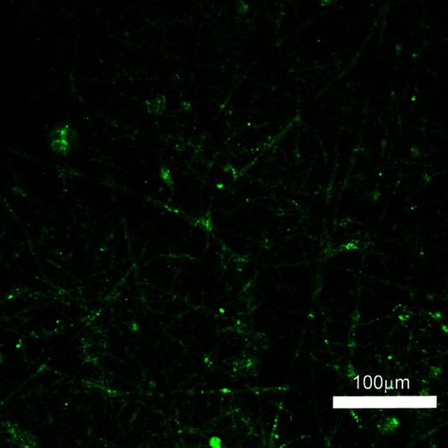
Figure 4 FTIR spectra of electrospun pure PLGA and lidocaine/hEGF-loaded PLGA nanofibers.
Abbreviations: FTIR- fourier-transform infrared spectroscopy; hEGF, human epidermal growth factor; PLGA, poly[(d,l)-lactide-co-glycolide]; FTIR, fourier-transform infrared spectroscopy.
![Figure 4 FTIR spectra of electrospun pure PLGA and lidocaine/hEGF-loaded PLGA nanofibers.Abbreviations: FTIR- fourier-transform infrared spectroscopy; hEGF, human epidermal growth factor; PLGA, poly[(d,l)-lactide-co-glycolide]; FTIR, fourier-transform infrared spectroscopy.](/cms/asset/031d0b64-c243-4761-b9a7-f6f80928610d/dijn_a_12190882_f0004_c.jpg)
Figure 5 Measured contact angles. (A) Pure PLGA nanofibers, 127.8, (B) lidocaine-incorporated nanofibers, 52.3, and (C) lidocaine/hEGF-loaded nanofibers, 60.1.
Abbreviations: hEGF, human epidermal growth factor; PLGA, poly[(d,l)-lactide-co-glycolide].
![Figure 5 Measured contact angles. (A) Pure PLGA nanofibers, 127.8, (B) lidocaine-incorporated nanofibers, 52.3, and (C) lidocaine/hEGF-loaded nanofibers, 60.1.Abbreviations: hEGF, human epidermal growth factor; PLGA, poly[(d,l)-lactide-co-glycolide].](/cms/asset/352a7e38-aa2a-4b97-bcc1-42bd9dd5bfbd/dijn_a_12190882_f0005_b.jpg)
Figure 6 Stress–strain curve of pure PLGA, lidocaine-incorporated, and lidocaine/hEGF-loaded sheath-core-structured nanofibers.
Abbreviations: hEGF, human epidermal growth factor; PLGA, poly[(d,l)-lactide-co-glycolide].
![Figure 6 Stress–strain curve of pure PLGA, lidocaine-incorporated, and lidocaine/hEGF-loaded sheath-core-structured nanofibers.Abbreviations: hEGF, human epidermal growth factor; PLGA, poly[(d,l)-lactide-co-glycolide].](/cms/asset/1d7a8fa0-3102-498e-a316-8633f4c840c9/dijn_a_12190882_f0006_c.jpg)
Figure 7 In vitro release patterns of anesthetics from the nanofibrous films.
Notes: (A) Daily release, (B) accumulated release.
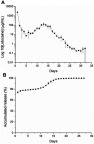
Figure 8 In vivo release of lidocaine from the nanofibrous films.
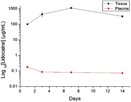
Figure 9 In vitro elution profiles of hEGF from the nanofibrous films.
Abbreviation: hEGF, human epidermal growth factor.
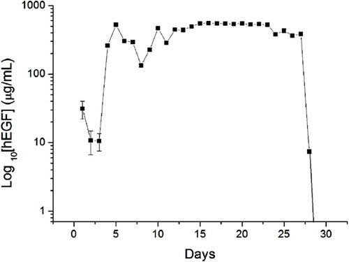
Figure 10 In vivo release of hEGF from the nanofibrous films.
Abbreviation: hEGF, human epidermal growth factor.
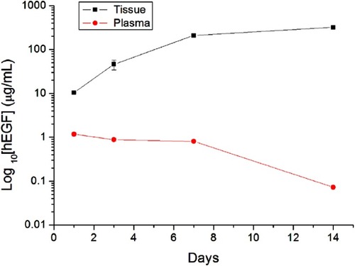
Figure 11 Cell viability of electrospun lidocaine/hEGF nanofibers (##P<0.01, **P<0.01).
Abbreviations: hEGF, human epidermal growth factor; EGF, epidermal growth factor.
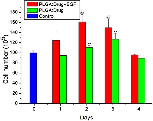
Figure 12 Activities in the rats.
Abbreviation: EGF, epidermal growth factor; PLGA, poly[(d,l)-lactide-co-glycolide].
![Figure 12 Activities in the rats.Abbreviation: EGF, epidermal growth factor; PLGA, poly[(d,l)-lactide-co-glycolide].](/cms/asset/851b2abe-deae-4e62-be7b-88f5c2a08abd/dijn_a_12190882_f0012_c.jpg)
Figure 13 No tissue adhesion was observed for the wounds implanted with the sheath-core membranes.
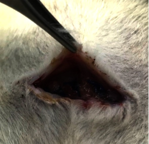
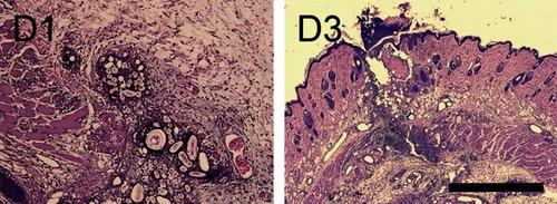
Figure 14 Wound healing in hEGF/lidocaine-incorporated nanofibers at post-surgical days 1 and 3 (scale bar: 1 mm). A foreign body reaction was noted at day 1 (D1), while fibroblastic proliferation and mixed infiltrates of lymphocytes and plasma cells were observed in the subcutis and muscular layer on day 3 (D3).
Abbreviation: hEGF, human epidermal growth factor.