Figures & data
Figure 1 (A) Variation of particle size and zeta potential as a function of CM ratio (SLN/Ce6 to CM protein, w/w). Inset image was the optical image of CM/SLN/Ce6 at the optimal w/w ratio, which provides direct evidence of successful Ce6 loading. (B) Western blot analysis of two proteins (AT1R and CXCR4) in CM and CM/SLN/Ce6. (C) TEM images of SLN/Ce6, CM and CM/SLN/Ce6. Scale bar: 100 nm.
Abbreviations: Ce6, chlorins e6; CM, cellular membranes; TEM, transmission electron microsope; SLN, silica nanoparticles.
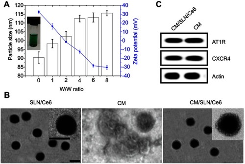
Figure 2 (A) Colloidal stability of CM/SLN/Ce6 in PBS (pH 7.4) and mouse plasma at 37°C for up to 48 hours. (B) Comparative fluorescentstability of CM/SLN/Ce6 and free Ce6.
Note: Data were shown as mean ±SD (N=3). *p<0.05.
Abbreviations: FL, fluorescent; Ce6, chlorins e6; CM, cellular membranes; SLN, silica nanoparticles.
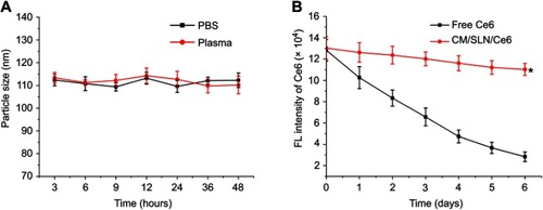
Figure 3 (A) Drug release profiles of Ce6 from the CM/SLN/Ce6 in release media under extracellular and intracellular condition of pHs (7.4 and 5.5). (B) Variation on absorbance of DPBF at 418 nm in the presence of CM/SLN/Ce6 (Ce6 concentration 0.1 mg/mL) after irradiation (1 W/cm2) for different time intervals. (B) Variation on absorbance of DPBF in the presence of different concentrations of CM/SLN/Ce6 (Ce6 concentration: 0.1–0.5 mg/mL) under irradiation for 60 seconds. (D) The in vitro ROS generation analysis of CM/SLN/Ce6. Main figure illustrated flow cytometric analysis of MFI in cells treated by different formulations after 5 minutes of laser irradiation (1 W/cm2). Inset images demonstrated the representing CLSM images of cells treated with different formulations.
Notes: Significant differences were vs CM/SLN/Ce6. Scale bar: 20 μm. Data were shown as mean ±SD (N=3). *p<0.05.
Abbreviations: Ce6, chlorins e6; CM, cellular membranes; SLN, silica nanoparticles; DPBF, 1,3-diphenylisobenzofuran; MFI, mean fluorescence intensity.
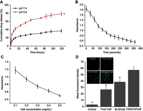
Figure 4 (A) Cytotoxicity of CM/SLN after 48 hours incubation with SGC7901 cells. (B) Cytotoxicity of free Ce6, SLN/Ce6 and CM/SLN/Ce6 (with laser irradiation) at different Ce6 concentrations against SGC7901 cells after 48 hours incubation. Inset images demonstrated the cleaved caspase-3, cytochrome C and bcl-2 levels in three groups (Ce6 concentration: 5 μg/mL).
Note: Data were shown as mean ±SD (N=3).
Abbreviations: Ce6, chlorins e6; CM, cellular membranes; SLN, silica nanoparticles.
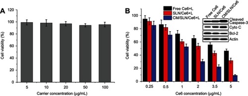
Figure 5 (A) Quantitative analysis of intracellular time-dependent uptake of different formulations in SGC7901 cells (pretreated with/without CM). Significant differences were vs groups pretreated without CM. (B) Representative CLSM images of uptake of different formulations in SGC7901 cells (pretreated with/without CM) at 4 hours postincubation. Scale bar: 20 μm. (C) In vivo real time distribution of SLN/ICG and CM/SLN/ICG at 6 and 12 hours postincubation. (D) Mean fluorescentintensity of dissected tumors and major organs of mice treated with SLN/ICG and CM/SLN/ICG at 48 hours postinjection.
Note: Data were expressed as mean ±SD (N=3).
Abbreviations: Ce6, chlorins e6; CM, cellular membranes; SLN, silica nanoparticles; CLSM, confocal laser scanning microscope; ICG, indocyanine green dye.
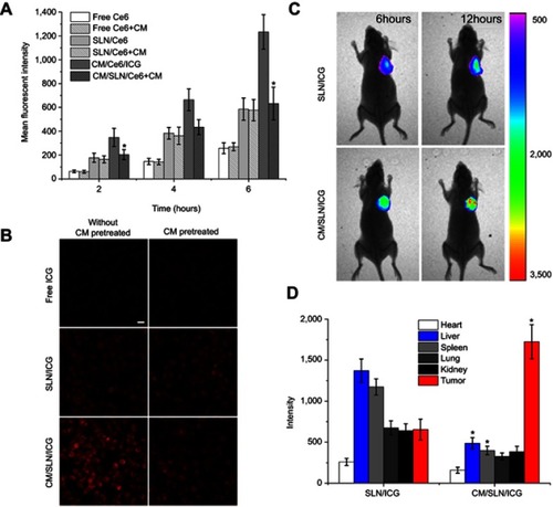
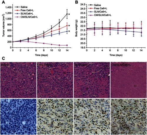
Figure 6 The tumor volume (A), body weight (B) and H&E/TUNEL staining (200×) of tumor tissue (C) analysis of SGC7901 tumor-bearing Balb/c nude mice after intravenous administration of different formulations.
Notes: Data were expressed as mean ±SD (N=6). Significant differences were vs CM/SLN/Ce6. *p<0.05.
Abbreviations: Ce6, chlorins e6; CM, cellular membranes; SLN, silica nanoparticles.