Figures & data
Figure 1 Preparation of the water extract of Gusuibu (WEG) and nanoparticles of the WEG (nWEG). Dry pieces of Gusuibu (A), Drynaria fortunei, were ground into a fine powder. WEG (B) and nWEG were prepared as described in Materials and Methods. The distribution of nWEG particles was analyzed using an atomic force microscope (C).
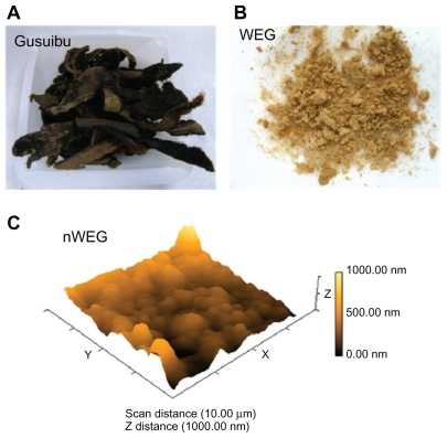
Figure 2 Effects of nanoparticles prepared from the water extract of Gusuibu (nWEG) on cell viability and apoptotic cells. Primary rat osteoblasts isolated from neonatal calvarias were exposed to 1, 10, 100, and 1000 μg/mL of nWEG for 72 hours. The viability of rat osteoblasts was assayed using a colorimetric method (A). Cell apoptosis was quantified by flow cytometry (B).
Note: Each value represents the mean ± SEM for n = 6.
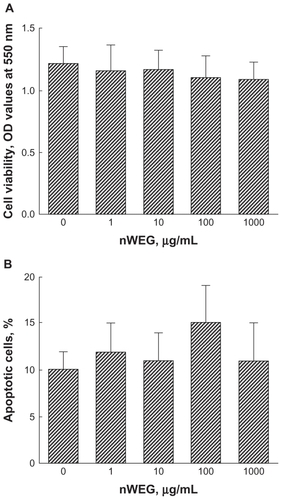
Figure 3 Effects of the water extract of Gusuibu (WEG) and nanoproducts of the WEG (nWEG) on cell viability. Primary rat osteoblasts isolated from neonatal calvarias were exposed to 1000 μg/mL of WEG (A) and nWEG (B) for 24, 48, and 72 hours. Cell viability was assayed according to a colorimetric method.
Notes: Each value represents the mean ± SEM for n = 6. *Indicates that a value significantly differs from the respective control, P < 0.05.
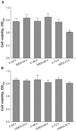
Figure 4 Effects of the water extract of Gusuibu (WEG) and nanoproducts of the WEG (nWEG) on DNA fragmentation and cell apoptosis. Primary rat osteoblasts isolated from neonatal calvarias were exposed to 1000 μg/mL of WEG and nWEG for 72 hours. DNA fragmentation was assayed using an enzyme-linked immunosorbent assay kit (A). Cell apoptosis was analyzed and quantified by flow cytometry (B, C). Sodium nitroprusside (SNP) was administered to rat osteoblasts as a positive control.
Notes: Each value represents the mean ± SEM for n = 6. *Indicate that a value significantly (P < 0.05) differs from control and WEG-treated groups, respectively.
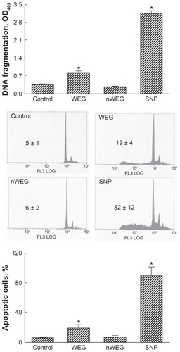
Figure 5 Effects of the water extract of Gusuibu (WEG) and nanoproducts of the WEG (nWEG) on hydrogen peroxide- (HP) and sodium nitroprusside (SNP)-induced cell insults. Primary rat osteoblasts isolated from neonatal calvarias were exposed to 100 μM HP, 10 μg/mL WEG, 10 μg/mL nWEG, and a combination of HP with WEG or nWEG for 24 hours. Cell viability (A), DNA fragmentation (B), and cell apoptosis (C) were quantified. SNP was administered to rat osteoblasts, and cell viability was assayed (D).
Note: Each value represents the mean ± SEM for n = 6. *,#,+Indicate that a value significantly (P < 0.05) differs from control, and HP- and WEG-treated groups, respectively.
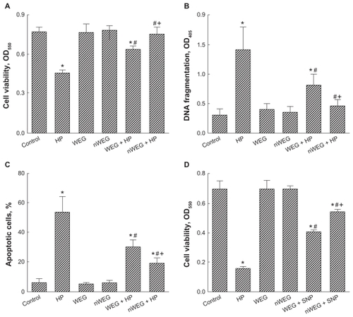
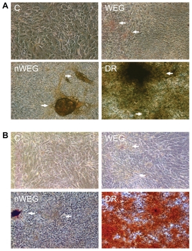
Figure 6 Effects of the water extract of Gusuibu (WEG) and nanoproducts of the WEG (nWEG) on osteoblast maturation. Primary rat osteoblasts isolated from neonatal calvarias were exposed to 10 μg/mL WEG, 10 μg/mL nWEG, or a differentiation agent, including dexamethasone, ascorbic acid, and β-glycerophosphate, for 21 days. Mineralized nodules were stained using the von Kossa (A) and Alizarin red S-staining protocols (B), and then photographed with a light microscope. 100×.