
Figures & data
Scheme 1 Hydroxyapatite-coated PLLA/PCL hierarchical nanofibrous scaffold fabricated via a one-pot thermally induced phase separation (TIPS) and electrochemical deposition (ED) technique.Abbreviations: HA, hydroxyapatite; PLLA, poly(l-lactic acid); PCL, poly(ε-caprolactone).
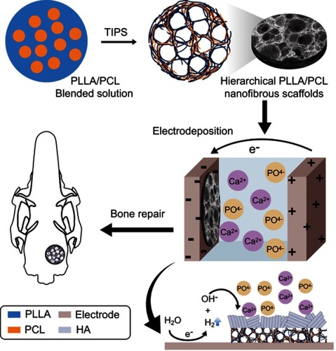
Figure 1 (A) Photo of PLLA/PCL scaffold. (B) SEM images of PLLA/PCL scaffold (C) a magnified portion of the image (B). (D) The pore size distribution of the PLLA/PCL scaffold. Abbreviations: SEM,scanning electron microscopy;PLLA, poly(l-lactic acid); PCL, poly(ε-caprolactone).
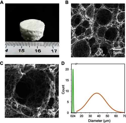
Figure 2 (A–C) SEM images of mineralized PLLA/PCL scaffold at 37°C and 2 V for: (A) 1 hr, (B) 2 hrs, (C) 3 hrs. (D–F) SEM images of mineralized PLLA/PCL scaffold at 37°C and 2 hrs for (A) 1 V, (B) 2 V, (C) 3 V. The insets are of magnified images, and the magnification of SEM figures and inlets is uniform. (G) Mass increase with different time. (H) Mass increase with different voltage. (I) XRD patterns of the corresponding mineralized PLLA/PCL scaffold.Abbreviations: V, voltage;SEM,scanning electron microscopy;XRD,X-ray diffraction;PLLA, poly(l-lactic acid); PCL, poly(ε-caprolactone).
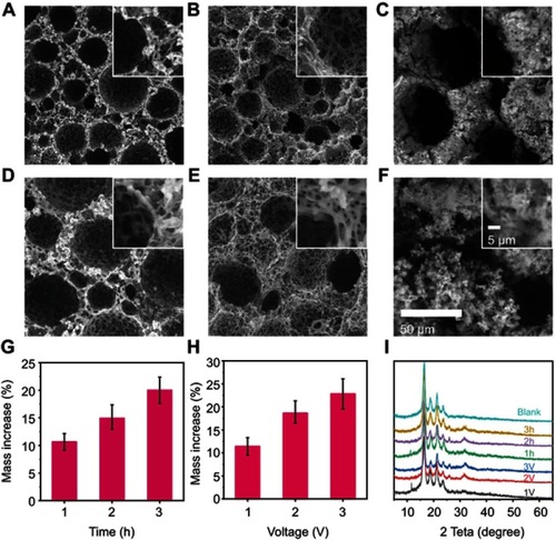
Figure S1 EDS mapping of the cross-section of the M-PLLA/PCL scaffold. (A) Ca elemental distribution. (B) P elemental distribution. (C) C elemental distribution. (D) The quantitative analysis of the elements on the scaffold.Abbreviations: EDS, electron dispersive spectrometry; PLLA, poly(l-lactic acid); PCL, poly(ε-caprolactone).
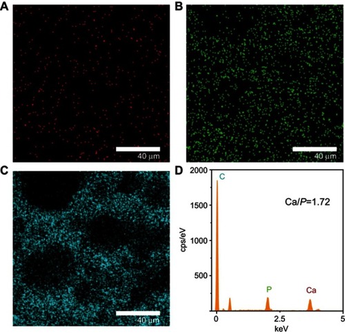
Figure 3 The rBMSCs adhered (A) and proliferated (B) on the scaffolds, as detected by CCK-8 test. (C) The SEM and (D) confocal tomography of the rBMSCs on the M-PLLA/PCL scaffold after 7 days culture.Abbreviations: rBMSCs, rat bone marrow stromal cells; CCK-8, Cell Counting Kit-8;SEM,Scanning electron microscopy;M-PLLA/PCL, hydroxyapatite- coated hierarchical PLLA/PCL nanofibrous scaffold;PLLA, poly(l-lactic acid); PCL, poly(ε-caprolactone).
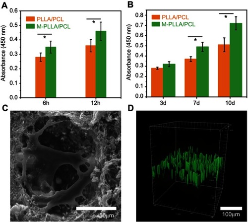
Figure 4 (A) ALP activity at designated time intervals during osteogenic induction; (B) relative expressions of Col1A1, OCN, RUNX2, and OPN by rBMSCs induced on different scaffolds for 2 weeks. *p<0.05 and **p<0.01.Abbreviations: rBMSCs, rat bone marrow stromal cells; PLLA, poly(l-lactic acid); PCL, poly(ε-caprolactone);M-PLLA/PCL,hydroxyapatite-coated hierarchical PLLA/PCL nanofibrous scaffold.
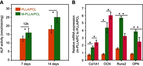
Figure 5 (A) Micro-CT evaluation (sagittal views and 3D surface rendering) of in vivo calvarial bone healing at 2, 4, and 12 weeks post-implantation for PLLA/PCL and M-PLLA/PCL. (B) The change of bone mineral density (BMD) after the scaffold implantation. (C) The change in bone volume (BV) after the scaffold implantation.(* p<0.05 and ** p<0.01)Abbreviations: rBMSCs, rat bone marrow stromal cells; PLLA, poly(l-lactic acid); PCL, poly(ε-caprolactone);M-PLLA/PCL,hydroxyapatite-coated hierarchical PLLA/PCL nanofibrous scaffold.
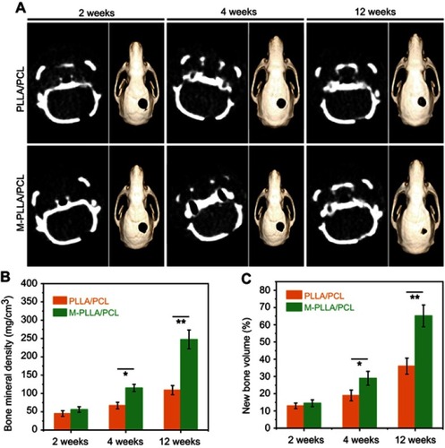
Figure 6 Histological analysis for in vivo calvaria defect repair. The magnification of figures and inlets is uniform.Abbreviations: PLLA, poly(l-lactic acid); PCL, poly(ε-caprolactone);M-PLLA/PCL,hydroxyapatite-coated hierarchical PLLA/PCL nanofibrous scaffold.
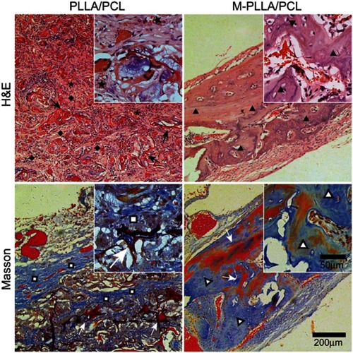
Table S1 Mechanical properties of the M-PLLA/PCL and PLLA/PCL (*p<0.05)