Figures & data
Table 1 Inhibitors with different functions used in the transport study and their concentrations
Figure 1 (A) Schematic illustration of ion-pairing complex formation between PMX and DCK, and self-assembled micelle formation of PMX/DCK-OP in the aqueous phase. (B) Transmission electron micrograph of PMX/DCK-OP micelles. Scale bar represents 200 nm.
Abbreviations: PMX, pemetrexed; DCK, Nα-deoxycholyl-L-lysyl-methylester; PMX/DCK, ion-pairing complex between PMX and DCK; PMX/DCK-OP, oral powder formulation of PMX/DCK.
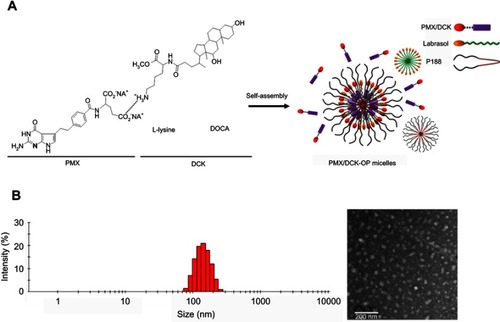
Table 2 Effective and apparent permeability of PMX, PMX/DCK, and PMX/DCK-OP
Figure 2 (A) Relative apparent permeability of PMX/DCK-OP across a Caco-2 cell monolayer after incubation with various transport inhibitors and (B) with or without EGTA in the presence or absence of all inhibitors except cyclosporine A.
Notes: Each value represents the mean ± standard deviation (n=6). **P<0.01, ***P<0.001 compared to the Papp of the untreated control group. ###P<0.001 compared to the apparent permeability of PMX/DCK-OP after treatment with all inhibitors except cyclosporine A in the absence of EGTA. $$$P<0.001 compared to the Papp of PMX/DCK-OP after treatment with EGTA in the presence of all inhibitors except cyclosporine A.
Abbreviations: PMX, pemetrexed; DCK, Nα-deoxycholyl-L-lysyl-methylester; PMX/DCK, ion-pairing complex between PMX and DCK; PMX/DCK-OP, oral powder formulation of PMX/DCK; Act D, actinomycin D; MβCD, methyl-β-cyclodextrin; ASBT; EGTA, ethylene glycol-bis-(β-aminoethyl ether)-N,N,N′,N′-tetraacetic acid; Papp, apparent permeability of PMX/DCK-OP across a Caco-2 cell monolayer.
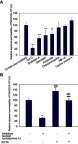
Figure 3 Schematic diagrams of possible specific and nonspecific transport pathways, and predicted intracellular trafficking and transcytosis of PMX/DCK-OP across an intestinal epithelium: (1) ASBT-mediated transport; (2) caveola/lipid raft-mediated endocytosis; (3) macropinocytosis; (4) paracellular transport; and (5) transcellular passive diffusion.
Abbreviations: PMX, pemetrexed; DCK, Nα-deoxycholyl-L-lysyl-methylester; PMX/DCK, ion-pairing complex between PMX and DCK; PMX/DCK-OP, oral powder formulation of PMX/DCK; ASBT, apical sodium-dependent bile acid transporter; IBABP, intestinal bile acid-binding protein; OSTα, organic solute transporter alpha; OSTβ organic solute transporter beta; ER, endoplasmic reticulum.
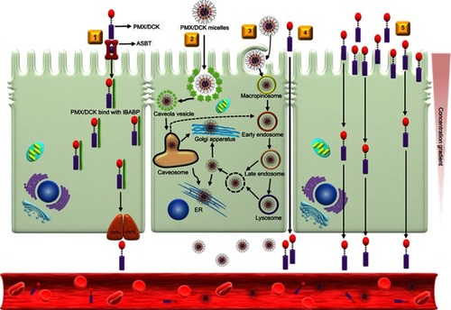
Table 3 Pharmacokinetic parameters of PMX in rats after intravenous injection of PMX and oral administration of PMX, PMX/DCK, or PMX/DCK-OP
Figure 4 Venous plasma concentration-time profiles of PMX after (A) single intravenous administration of PMX (5 mg/kg) and (B) oral administration of PMX (25 mg/kg), PMX/DCK (equivalent to 25 mg/kg of PMX), or PMX/DCK-OP (equivalent to 25 mg/kg of PMX) in aqueous solution to rats.
Notes: Each value represents the mean ± standard deviation (n=4 for each group).
Abbreviations: PMX, pemetrexed; DCK, Nα-deoxycholyl-L-lysyl-methylester; PMX/DCK, ion-pairing complex between PMX and DCK; PMX/DCK-OP, oral powder formulation of PMX/DCK.
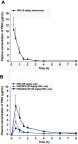
Figure 5 Efficacy of in vivo tumor growth inhibitory effects in B16F10 tumor-bearing mice after biweekly intravenous administration of 20 mg/kg PMX (PMX-IV), intraperitoneal administration of 10 mg/kg anti-PD1 (Anti-PD1) once every 3 days, once-daily oral administration of PMX/DCK-OP as 20 mg/kg PMX (PMX/DCK-OP), and combined treatment with once-daily oral administration of PMX/DCK-OP as 20 mg/kg PMX and intraperitoneal administration of 10 mg/kg anti-PD1 (PMX/DCK-OP+Anti-PD1) once every 3 days for 14 days. (A) Tumor volume in mice (**P<0.01, ***P<0.001). (B) Variation of body weight in mice during treatment. (C) Tumor weight in B16F10 tumor-bearing mice (**P<0.01, ***P<0.001 compared to the control group). (D) Photographs of tumors isolated from each group on day 15. Scale bar represents 10 mm.
Notes: Statistics: one-way ANOVA followed by Tukey’s multiple-comparison test. Each value represents the mean ± standard deviation (n=10 for each group).
Abbreviations: PMX, pemetrexed; DCK, Nα-deoxycholyl-L-lysyl-methylester; PMX/DCK, ion-pairing complex between PMX and DCK; PMX/DCK-OP, oral powder formulation of PMX/DCK.
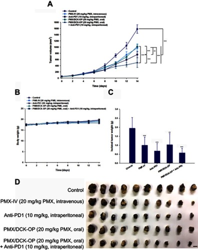
Figure 6 Representative cross-sectional images of isolated tumor tissues stained with (A) anti-CD31 antibody for microvessels (brown), (B) proliferating cell nuclear antigen (PCNA) for proliferating cells (brown), and (C) fluorescent terminal deoxynucleotidyl transferase-mediated dUPT nick end labeling (TUNEL) for apoptosis (green fluorescence) in the tumor tissues taken 14 days after treatment with various modes.
Notes: The treatment modes were biweekly intravenous administration of 20 mg/kg PMX (PMX-IV), intraperitoneal administration of 10 mg/kg anti-PD1 (Anti-PD1) once every 3 days, once-daily oral administration of PMX/DCK-OP as 20 mg/kg of PMX (PMX/DCK-OP), and combined once-daily oral administration of PMX/DCK-OP as 20 mg/kg of PMX with once every 3 days intraperitoneal administration of 10 mg/kg anti-PD1 (PMX/DCK-OP + Anti-PD1). Scale bars represent 50 μm for anti-CD31 antibody and PCNA staining, and 20 μm for TUNEL staining, respectively.
Abbreviations: PMX, pemetrexed; DCK, Nα-deoxycholyl-L-lysyl-methylester; PMX/DCK, ion-pairing complex between PMX and DCK; PMX/DCK-OP, oral powder formulation of PMX/DCK.
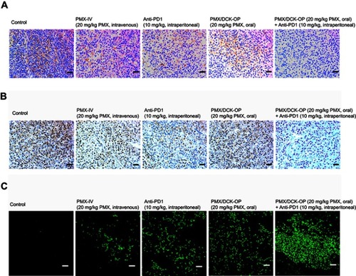
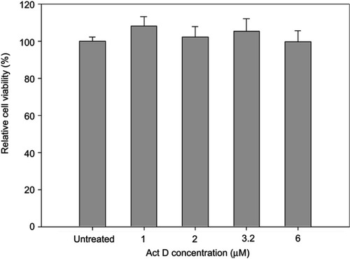
Figure S1 In vitro cytotoxic effect of Act D on Caco-2 cells.
Notes: Cell viability was measured using WST-8 and the growth of Caco-2 cells compared to the untreated control group. All data are expressed as means ± standard deviation (n=6).
Abbreviation: Act D, actinomycin D.