Figures & data
Figure 1 Protocol of iron oxide nanoparticle administration and OVA sensitization. BALB/c mice were randomly divided into the following groups (3–5 animals/group): naïve (NA), ovalbumin-sensitized (OVA), vehicle-treated and OVA-sensitized (VH), and iron oxide nanoparticle-treated and OVA-sensitized. Except for NA and OVA groups, the mice were administered intravenously with a single dose of iron oxide nanoparticles (10, 30 and 60 mg Fe/kg; 0.2 mL/mouse) and/or vehicle (VH; saline; 0.2 mL/mouse). Except for the NA group, the mice were sensitized 1 hour after drug administration with ovalbumin (OVA) by intraperitoneal injection using a sensitization solution containing 100 μg OVA plus 1 mg alum (as adjuvant) in saline (250 μL/mouse). Mice were sacrificed 7 days after OVA sensitization, and the spleen and blood were harvested for further experimentation.
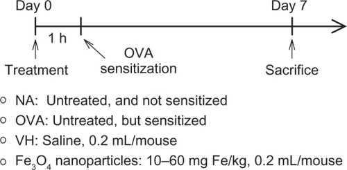
Figure 2 Attenuation by iron oxide nanoparticles of the serum production of OVA-specific IgG1 and IgG2a. Mice were treated with iron oxide nanoparticles and sensitized with OVA as depicted in . The serum levels of OVA-specific IgG1 and IgG2a were measured by ELISA.
Note: Data are expressed as the mean ± SE of 9–15 samples pooled from 3 independent experiments. *P < 0.05 compared to the VH group.
Abbreviations: OVA, ovalbumin; ELISA, enzyme-linked immunosorbent assay; SE, standard error; VH, vehicle treated group; OD, optical density.
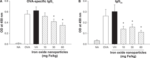
Table 1 Exposure to iron oxide nanoparticles did not affect splenocyte cellularity
Figure 3 Attenuation by iron oxide nanoparticles of antigen-induced production of IFN-γ and IL-4 by splenocytes. Mice were treated with iron oxide nanoparticles and sensitized with OVA as depicted in . The splenocytes (5 × 10⁶ cells/mL) isolated from each group of mice were re-stimulated with OVA (50 μg/mL) in culture for 72 hours to induce cytokine production. The levels of (A) IFN-γ and (B) IL-4 in the supernatants were measured by ELISA.
Notes: Data are expressed as the mean ± SE of quadruplicate cultures. *P < 0.05 compared to the VH control. Results are a representative of 3 independent experiments.
Abbreviations: OVA, ovalbumin; ELISA, enzyme-linked immunosorbent assay; SE, standard error; VH, vehicle treated group.
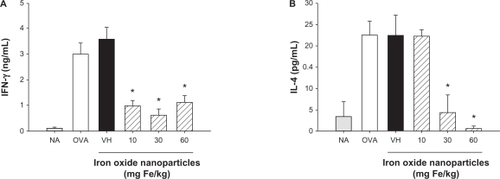
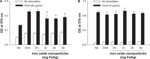
Figure 4 Effect of iron oxide nanoparticles on the viability of splenocytes. Mice were treated with iron oxide nanoparticles and sensitized with OVA as depicted in . The splenocytes (5 × 10⁶ cells/mL) isolated from each group of mice were cultured in the absence or presence of OVA (50 μg/mL) for 72 hours or ConA (5 μg/mL) for 48 hours, and the viability of splenocytes was determined by an MTT assay.
Notes: Data are expressed as the mean ± SE of quadruplicate cultures. *P < 0.05 as compared with the matched VH group. Results are representative of 3 independent experiments.
Abbreviations: OVA, ovalbumin; ConA, concanavalin A; MTT, 3-(4,5-dimethylthiazol-2-yl)-2,5-diphenyl-tetrazolium bromide; SE, standard error; VH, vehicle treated group; OD, optical density.