Figures & data
Figure 1 Schematic diagram for the synthesis of SPIO/DSPE-PEG5k-(Bom&Cy5).
Abbreviations: SPIO, superparamagnetic iron oxide; NHS, N-hydroxysuccinimide.
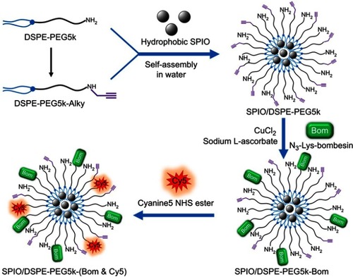
Figure 2 Nanomicelle characterization.
Notes: (A) A TEM image of Bom-targeted nanomicelles (scale bar =100 nm). (B) Size distribution of Bom-targeted (blue) and nontargeted (red) nanomicelles in aqueous solution measured by DLS.
Abbreviations: TEM, transmission electron microscopy; DLS, dynamic light scattering.
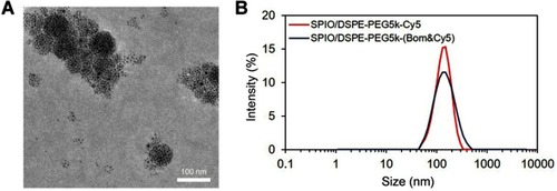
Figure 3 Relaxivity and magnetization measurement.
Notes: (A) T2-weighted images of SPIO/DSPE-PEG5k-(Bom&Cy5) nanomicelle samples at different iron concentrations (3.0 T, spin-echo sequence: TR =5000 ms, TE =12 ms). (B) Chart of the change in 1/T2 values with Fe concentration. (C) Hysteresis loops of the SPIO nanoparticles (black) and SPIO/DSPE-PEG5k-(Bom&Cy5) nanomicelles (red) measured at 300 K.
Abbreviation: SPIO, superparamagnetic iron oxide.
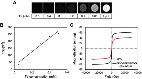
Figure 4 Cytotoxicity and in vivo toxicology.
Notes: (A) In vitro cell viability of HUVEC, L02 and HEK-293 cells after incubation with various concentrations of SPIO/DSPE-PEG5k-(Bom&Cy5) nanomicelles for 24 hrs. (B) Serum biochemistry data on ALT, AST, BUN and CREA. (C) Micrographs of H&E-stained organ slices (heart, lung, liver, spleen and kidney) from mice 3 days, 7 days and 21 days after intravenous injection of SPIO/DSPE-PEG5k-(Bom&Cy5) nanomicelles (H&E staining, 40×). Scale bar =50 µm.
Abbreviations: SPIO, superparamagnetic iron oxide; ALT, alanine aminotransferase; AST, aspartate aminotransferase; BUN, blood urea nitrogen; CREA, creatinine.
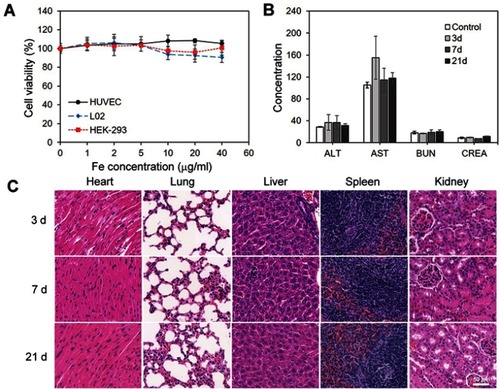
Figure 5 Targeting performance in cells.
Notes: Fluorescence microscopy of MDA-MB-231 cells incubated with Bom-targeted nanomicelles (1st line), nontargeted nanomicelles (2nd line) and Bom-targeted nanomicelles with free Bom (3rd line). From left to right are DAPI-labeled nuclei, Cy5-labeled nanomicelles and their overlay (scale bar =50 µm).
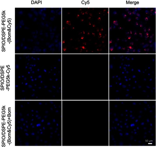
Figure 6 In vivo dual-modality imaging performance.
Notes: (A) MR images (3.0 T, T2-weighted fast spin echo sequence: TR =4000 ms, TE =66 ms, FOV =50 mm × 50 mm, slice thickness =1 mm) and (B) NSI of an MDA-MB-231 mouse xenograft tumor at different times after the intravenous injection of Bom-targeted nanomicelles and nontargeted nanomicelles. NSI=SItumor/SIwater phantom. (C) NIRF images and (D) average radiant efficiencies (p/s/cm2/sr)/(μW/cm2) at different times after the injection of Bom-targeted or nontargeted nanomicelles. The red arrows indicate tumors.
Abbreviations: NSI, normalized signal intensity; NIRF, near-infrared fluorescence.
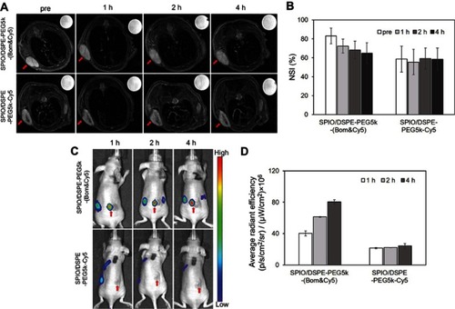
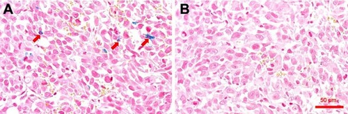
Figure 7 Prussian blue staining of tumor sections.
Notes: Prussian blue-stained tumor tissue section from mice treated with SPIO/DSPE-PEG5k-(Bom&Cy5) nanomicelles (A) and SPIO/DSPE-PEG5k-Cy5 nanomicelles (B), respectively. The red arrows indicate SPIO nanoparticles. Scale bar =50 µm.
Abbreviation: SPIO, superparamagnetic iron oxide.