Figures & data
Figure 1 The scheme of nanocomposite/photodynamic chemotherapy in tumor tissues and cells.
Abbreviations: Ce6, chlorin e6; DOX, doxorubicin hydrochloride.
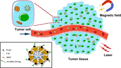
Figure 2 The structural characterization of the samples.
Notes: (A) SEM image of MSN. (B, C) TEM images of MSN. (D, E, F) TEM images of the Fe3O4 nanocrystals, MMSN, and nanocomposite. (G) Particle size of the MSN determined by DLS. (H) Zeta potential of the MSN determined by DLS.
Abbreviations: TEM, transmission electron microscope; SEM, scanning electron microscope; DLS, dynamic light scattering; MMSN, magnetic mesoporous silica nanoparticle.
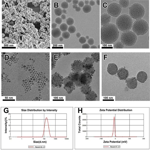
Figure 3 Characterization of the nanocomposites.
Notes: (A) Nitrogen absorption/desorption isotherms of MSN. Inset: the pore size distributions of MSN. (B) Hysteresis loop of samples normalized to the mass by VSM. Inset: photographs of a nanocomposite aqueous solution without (left) and with (right) magnetic field. (C) Near-infrared spectrogram of the samples. (D) Singlet-oxygen production of the samples in DMF.
Abbreviations: MSN, mesoporous silica nanoparticle; DMF, N,N-dimethylformamide; NIB, neodymium, iron, and boron magnet.
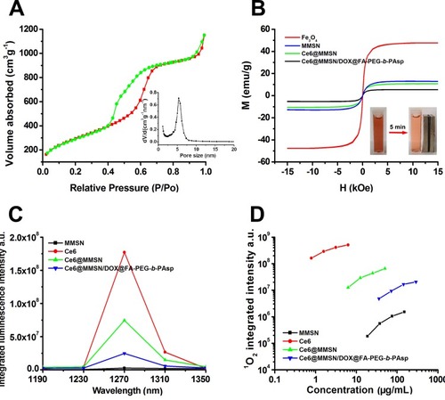
Figure 4 DOX release performance of the nanoparticles.
Notes: (A) UV absorption spectra of free DOX. (B) pH-responsive release profiles of the DOX in vitro.
Abbreviations: DOX, doxorubicin hydrochloride; MMSN, magnetic mesoporous silica nanoparticle.
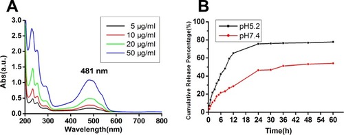
Figure 5 Cellular uptake of nanoparticles.
Notes: (A) Fluorescence images of MCF-7/ADR cells after 2-h incubation with DOX (a), Ce6@MMSN/DOX@PEG-b-PAsp (b), and Ce6@MMSN/DOX @FA-PEG-b-PAsp (c). (B) Biological TEM images of MCF-7/ADR cells treated with nanocomposites for 2 h. The circles and arrows indicate the locations of the nanoparticles. (C) The fluorescence intensity of MCF-7/ADR cells cultured under different conditions (right), C1, blank cell; C2, MSN; C3, nanocomposites without magnetic field; C4, nanocomposites within magnetic field; photographs of culture dishes containing MCF-7/ADR cells (left).
Abbreviations: TEM, transmission electron microscope; DAPI, 4′,6-diamidinio-2-phenylindole; DOX, doxorubicin hydrochloride; Ce6, chlorin e6.
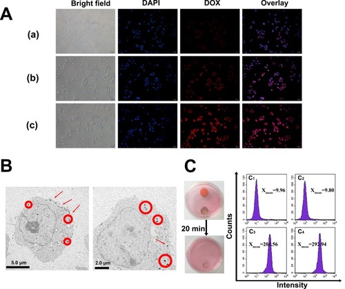
Figure 6 The therapeutic effect of the nanoparticles.
Notes: (A) Relative viabilities of MCF-7/ADR cells treated with different concentrations of free DOX and nanocomposites with or without light irradiation. (B) Half-maximal inhibition curve of MCF-7/ADR (a) and MCF-7 cells (b) treated with various concentrations of free DOX and nanocomposites (mean ± SD, n=3; *P<0.05, **P<0.01).
Abbreviations: DOX, doxorubicin hydrochloride; Ce6, chlorin e6; MMSN, magnetic mesoporous silica nanoparticle.
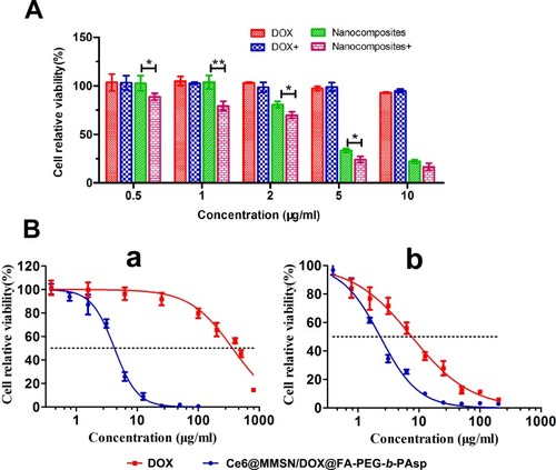
Figure 7 Results of the apoptosis assay and Transwell experiment of the nanocomposites in vitro.
Notes: (A) The apoptosis of MCF-7/ADR cells treated with MSN (a), free DOX (b) and nanocomposites (c) for 24 h. (B) Effects of nanocomposites on the invasion and motility of MCF-7/ADR cells. The cells were cultured with the indicated concentrations of nanocomposites. The cells were stained with crystal violet.
Abbreviations: FITC, fluorescein isothiocyanate; PI, propidium iodide; DOX, doxorubicin hydrochloride; MSN, mesoporous silica nanoparticle.
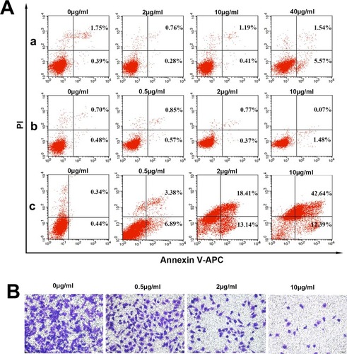
Figure 8 Magnetic targeting and histological analysis.
Notes: (A) T1-weighted MRI images of mice: control, postinjection (A1), and 2-h postinjection with NIB magnet for 30 min (A2). (B) Prussian blue-stained images; the circles and arrows indicate the locations of nanocomposites in the tumor site. (C) The histological characteristics of the main organs after treatment with 1 mg/mL MSN (a) and nanocomposites (b) once a day for 5 days (×200).
Abbreviations: MRI, magnetic resonance image; MMSN, magnetic mesoporous silica nanoparticle; NIB, neodymium, iron and boron magnet; MSN, mesoporous silica nanoparticle; H&E, hematoxylin and eosin.
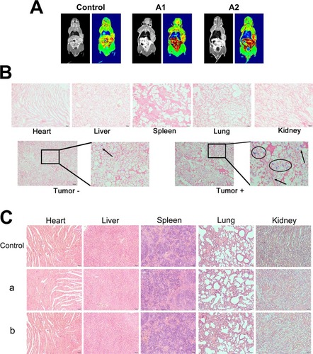
Table 1 Serum Biochemical Analysis Of The Liver Function In Nude Mice After Intravenous Treatments With 1 mg/mL MSN And Nanocomposites Once A Day For 5 Days. (n=5)
Table 2 Serum Biochemical Analysis Of The Renal Function In Nude Mice After Intravenous Treatments With 1 mg/mL MSN And Nanocomposites Once A Day For 5 Days. (n=5)
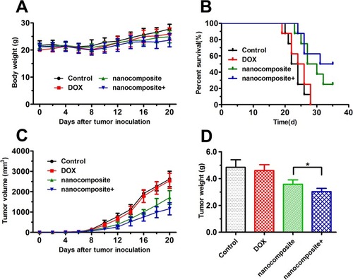
Figure 9 Antitumor experiments in vivo.
Notes: (A) The body weight changes of BALB/c nude mice during the therapeutic period. (B) Survival rate of MCF-7/ADR tumor-bearing BALB/c nude mice. (C) The tumor volume evolution of mice in different groups during the therapeutic period. (D) The weight of the excised tumor tissues from all groups. The data are expressed as the mean ± SD (n = 8). *P < 0.05.
Abbreviations: DOX, doxorubicin hydrochloride; MMSN, magnetic mesoporous silica nanoparticle; NIB, neodymium, iron and boron magnet.