Figures & data
Figure 1 Schematic illustration of the procedures for the synthesis of chitosan-doxorubicin conjugate.
Abbreviations: EDC, 1-ethyl-3-(3-dimethyl amino-propyl) carbodiimide hydrochloride; NHS, N-hydroxysuccinimide.
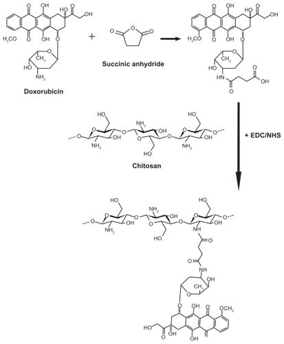
Table 1 Drug content and conjugation efficiency of different chitosan-doxorubicin conjugates
Figure 2 (A) Fourier transform infrared spectrum of doxorubicin and succinyl doxorubicin. (B) 1H nuclear magnetic resonance spectrum of doxorubicin and succinyl doxorubicin. (C) 1H nuclear magnetic resonance spectrum of chitosan and chitosan-doxorubicin conjugate.
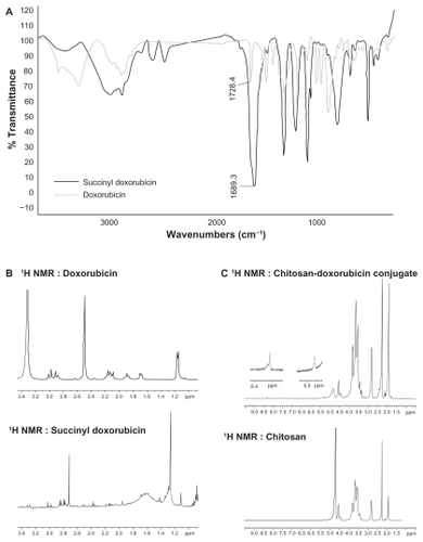
Figure 3 Differential scanning calorimetry thermograms of chitosan, doxorubicin and chitosan-doxorubicin conjugate (CS-DOX-2).
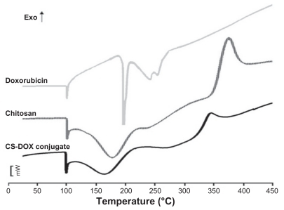
Figure 4 Gel permeation chromatogram of (A) chitosan and (B) chitosan-doxorubicin conjugates.
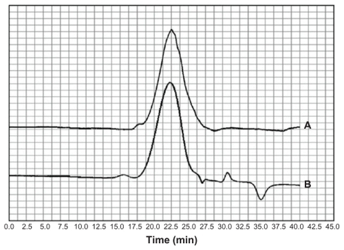
Table 2 Size, zeta potential, and polydispersity index of nanoaggregates prepared from different chitosan-doxorubicin conjugates
Figure 5 Effect of chitosan-doxorubicin conjugate concentration on hydrodynamic diameter of nanoaggregates.
Abbreviation: CS-DOX, chitosan-doxorubicin conjugate.
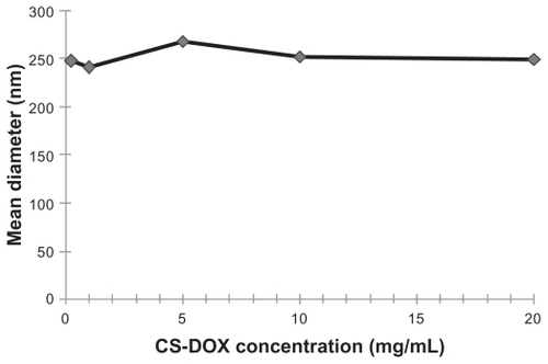
Figure 6 (A) Scanning electron micrographs of chitosan-doxorubicin conjugate (CS-DOX-2) nanoparticles and (B) transmission electron micrographs of CS-DOX- 2 nanoparticles.
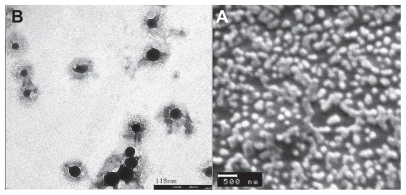
Figure 7 Isothermal titration calorimetry thermograms for interactions of protein A with (A) free antibody and (B) antibody attached to nanoparticles.
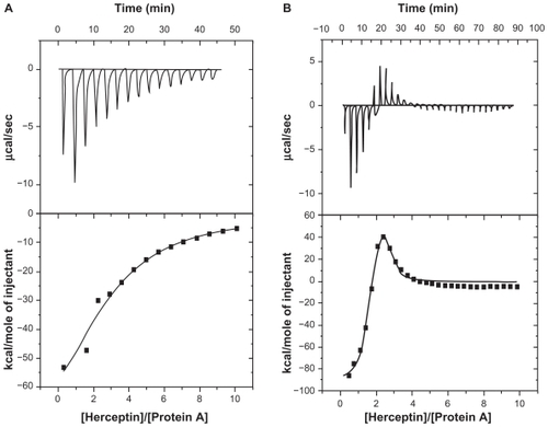
Table 3 Interaction thermodynamic parameters of protein A with free and attached antibody
Figure 8 Effect of doxorubicin, chitosan-doxorubicin conjugated nanoparticles, and trastuzumab-decorated chitosan-doxorubicin conjugated nanoparticles at different concentrations on viability of SKOV-3 cell line.
Abbreviations: CS, chitosan; DOX, doxorubicin; NP, nanoparticles.
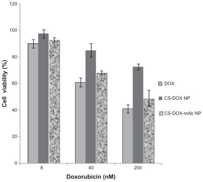
Figure 9 Fluorescence microscopy of cellular uptake of (A) trastuzumab-decorated chitosan-doxorubicin conjugated nanoparticles by SKOV-3 cell line, (B) trastuzumab-decorated chitosan-doxorubicin conjugated nanoparticles by MCF-7 cell line, (C) chitosan-doxorubicin conjugated nanoparticles by SKOV-3 cell line, and (D) chitosan-doxorubicin conjugated nanoparticles by MCF-7 cell line. (E) SKOV-3 cells and (F) MCF-7 cells in cell culture medium served as negative controls.
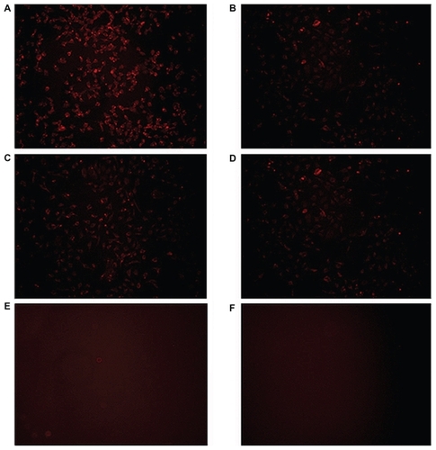
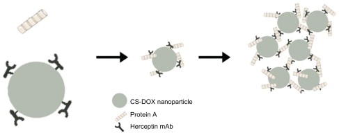
Figure 10 Schematic illustration of self-assembly of chitosan-doxorubicin conjugates into nanoparticles.
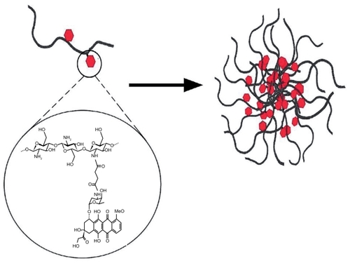
Figure 11 Interaction of protein A with antibodies attached to the nanoparticle surface.
Abbreviation: CS-DOX, chitosan-doxorubicin conjugate.