Figures & data
Table 1 Average diameter size of TiO2 nanoparticles bare, after functionalization and LbL deposition determined from TEM images
Figure 1 Example of transmission electron microscopy images of (A) bare TiO2 nanoparticles, (B) amino functionalized (TiO2-NH2) nanoparticles and (C) LbL coated (Q10). Bar represents 100 nm. (D) Thermograms of different LbL-DEX-coated Ti-O-NH2 substrate.
Abbreviations: DEX, dexamethasone; LbL, layer-by-layer.
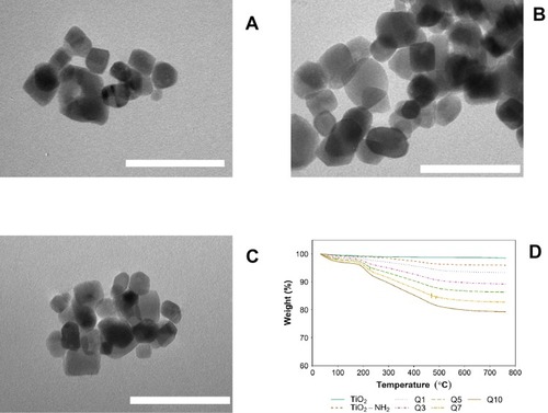
Table 2 Percentage of organic material in multi-layered DEX-LbL-loaded Ti-O-NH2 surface after addition of various quadruple layers (Q1, Q3, Q5, Q7 and Q10)
Table 3 DEX loading on multi-layered LbL-coated Ti-O-NH2 nanoparticles after addition of various quadruple layers (Q1, Q3, Q5, Q7 and Q10)
Figure 2 Cumulative release of DEX from LbL-Ti-O-NH2 surfaces at pH=5 (A) and pH=7.3 (B) for different number of quadruple layers (Q1, Q3, Q5, Q7 and Q10).
Abbreviations: PBAE, poly-beta-amino-ester; DEX-P, dexamethasone phosphate.
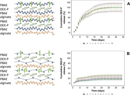
Figure 3 Mitochondrial activity of activated (A) and non-activated (B) THP-1 cells exposed to media containing DEX-P or elutes from DEX released from LbL assembly for 6 and 24 hrs. LPS concentration of 1 µg/mL.
Abbreviations: LPS, lipopolysaccharides; LbL, layer-by-layer; DEX, dexamethasone; DEX-P, dexamethasone phosphate.
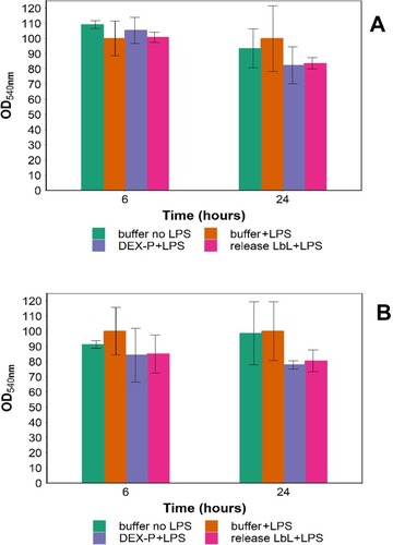
Figure 4 IL-6 expression of activated (A) and non-activated (B); TNFα expression of activated (C) and nonactivated (D) THP-1 cells post-exposure to media containing DEX-P or elutes from DEX released from LbL assembly for 6 and 24 hrs. LPS concentration of 1 µg/mL was used. (* represents significant differences p<0.05)
Abbreviations: DEX-P, dexamethasone phosphate; LbL, layer-by-layer; DEX, dexamethasone; LPS, lipopolysaccharides; TNFα, tumor necrosis factor alpha.
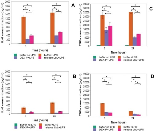
Figure 5 Actin staining epifluorescent images of human macrophages (A) LPS- DEX-; (B) LPS+ DEX-; (C) LPS+ DEX-P and (D) LPS+ DEX from release buffer after 24-hr exposure assessed by confocal microscopy. Actin rings and nuclei of cells were stained with phalloidin-FITC and DAPI, respectively; arrows indicate pseudopods. Bar corresponds to 20 µm.
Abbreviations: LPS, lipopolysaccharides; DEX, dexamethasone; DEX-P, dexamethasone phosphate.
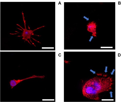
Figure 6 Mitochondrial activity of Saos-2 (A) and fibroblasts (B) cells exposed to media containing DEX-P or elutes from LbL assembly for 1, 2 and 3 days expressed as % of uncoated nanoparticles. (* represents significant differences compared to release from uncoated nanoparticles p<0.05).
Abbreviations: DEX-P, dexamethasone phosphate; LbL, layer-by-layer.
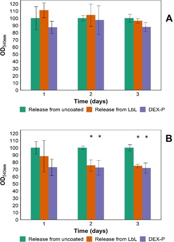
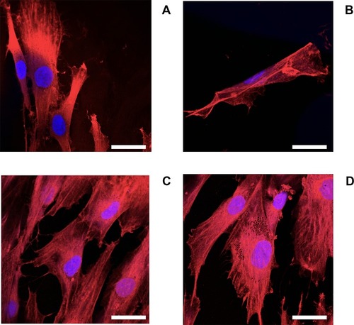
Figure 7 Actin staining epifluorescent images of human osteoblast (Saos-2) (A) no DEX; (B) LPS; (C) LPS+ DEX-P and (D) DEX from release buffer after 24-hr exposure assessed by confocal microscopy. Actin rings and nuclei of cells were stained with phalloidin-FITC and DAPI, respectively. Bar corresponds to 20 µm.
Abbreviations: LPS, lipopolysaccharides; DEX, dexamethasone; DEX-P, dexamethasone phosphate.
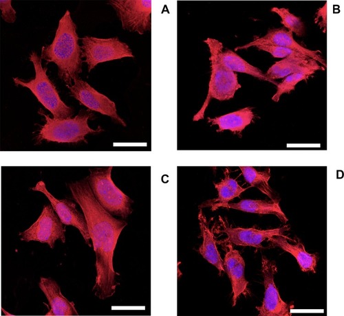
Figure 8 Actin staining epifluorescent images of human fibroblasts (A) no DEX; (B) LPS; (C) LPS+ DEX-P and (D) DEX from release buffer after 24-hr exposure assessed by confocal microscopy. Actin rings and nuclei of cells were stained with phalloidin-FITC and DAPI, respectively. Bar corresponds to 20 µm.
Abbreviations: LPS, lipopolysaccharides; DEX, dexamethasone; DEX-P, dexamethasone phosphate.