Figures & data
Figure 1 Structure and drug release mechanism of the PCC liposomes.
Abbreviations: PCC, photo-responsive Camellia sapogenin derivative cationic; SPC, soybean phosphatidylcholine; Chol, cholesterol; CMC, carboxymethyl chitosan; SPE, phosphatidyl ethanolamine; CTAB, cetrimonium bromide; CSD, Camellia sapogenin derivative.
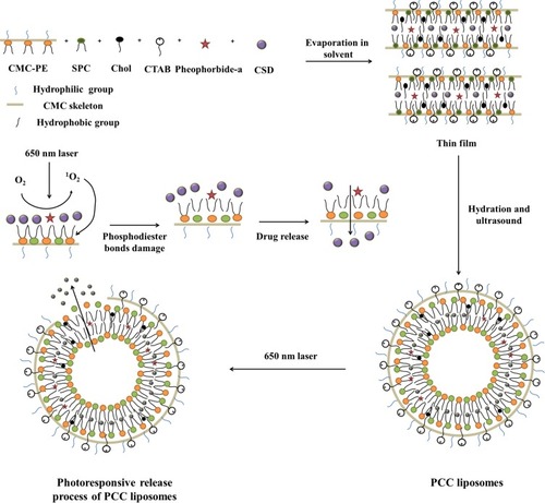
Figure 2 Synthetic route of Camellia sapogenin derivative.
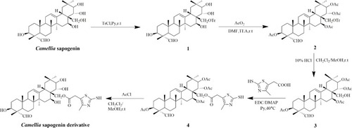
Figure 3 Synthetic route of PE-MAH and CMC-PE.
Abbreviations: CMC, carboxymethyl chitosan; MAH, maleic anhydride; PE, phosphatidyl ethanolamine.
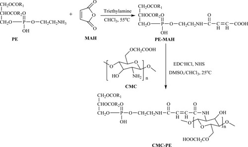
Figure 4 FTIR spectra of PE (A), PE-MAH (B) and CMC-PE (C).
Abbreviations: CMC, carboxymethyl chitosan; MAH, maleic anhydride; PE, phosphatidyl ethanolamine.
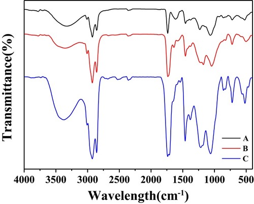
Figure 5 1H-NMR spectra of PE-MAH (A) and CMC-PE (B).
Abbreviations: CMC, carboxymethyl chitosan; MAH, maleic anhydride; PE, phosphatidyl ethanolamine.

Figure 6 Size distribution and zeta potential of the PCC liposomes.
Abbreviations: PCC, photo-responsive Camellia sapogenin derivative cationic.
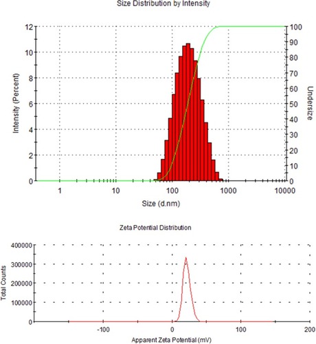
Figure 7 Fluorescence photographs of the PCC liposomes under white light (A) and 415 nm light excitation (B). TEM images of PCC liposomes. Scale bar = 1 μm (C) and 200 nm (D).
Notes: Red arrows represent pheophorbide-a in the outer shell and white arrows represent Camellia sapogenin derivative in the inner core of PCC liposomes.
Abbreviations: PCC, photo-responsive Camellia sapogenin derivative cationic; TEM, transmission electron microscopy.
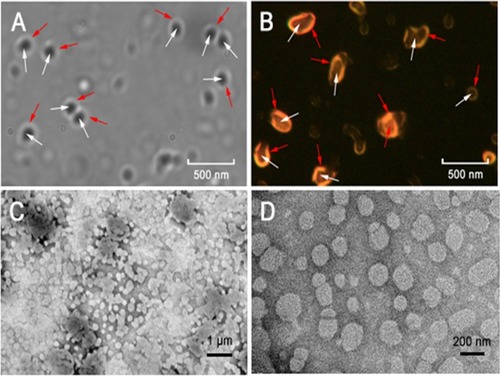
Figure 8 Changes of average size, zeta potential and encapsulation efficiency of the PCC liposomes stored at 4°C for 8 weeks.
Abbreviation: PCC, photo-responsive Camellia sapogenin derivative cationic.
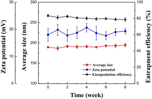
Figure 9 Retention rate of CSD in the PCC liposomes and contrast liposomes (without CMC-PE) in SGF and SIF.
Abbreviations: CSD, Camellia sapogenin derivative; PCC, photo-responsive Camellia sapogenin derivative cationic; SGF, simulated gastric fluid; SIF, simulated intestinal fluid; CMC, carboxymethyl chitosan; PE, phosphatidyl ethanolamine.
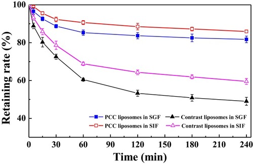
Figure 10 The UV-Vis and FL spectra of the PCC liposomes, normal blank liposomes and pheophorbide-a.
Abbreviations: PCC, photo-responsive Camellia sapogenin derivative cationic; UV-Vis, ultraviolet-visible; FL, fluorescence.
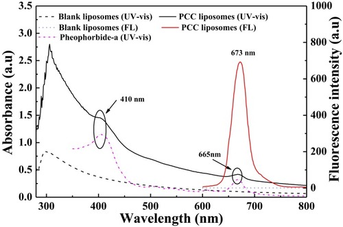
Figure 11 Cumulative release curves of CSD from the PCC liposomes at different illumination times (10, 20, 40, 60 mins).
Abbreviations: PCC, photo-responsive Camellia sapogenin derivative cationic; CSD, Camellia sapogenin derivative.
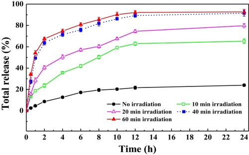
Figure 12 Hemolysis percentage of the PCC liposomes, blank cation liposomes, normal liposomes and CSD.
Abbreviations: PCC, photo-responsive Camellia sapogenin derivative cationic; CSD, Camellia sapogenin derivative.
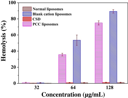
Figure 13 Morphology of rRBC after treatment with 1% triton (A), normal blank liposomes (B), blank cationic liposomes (C) and PCC liposomes (D) observed by inverted fluorescence microscope.
Abbreviations: PCC, photo-responsive Camellia sapogenin derivative cationic; rRBC, rabbit red blood cells.
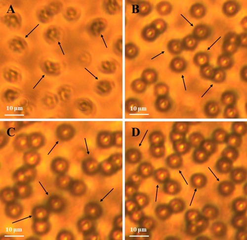
Figure 14 Cytotoxicity of the PCC liposomes, normal blank liposomes, blank cationic liposomes on HeLa cells after 24 hrs of incubation.
Abbreviation: PCC, photo-responsive Camellia sapogenin derivative cationic.
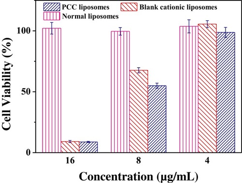
Table 1 Antibacterial Activity Of The Liposomes (unit: μg/mL)
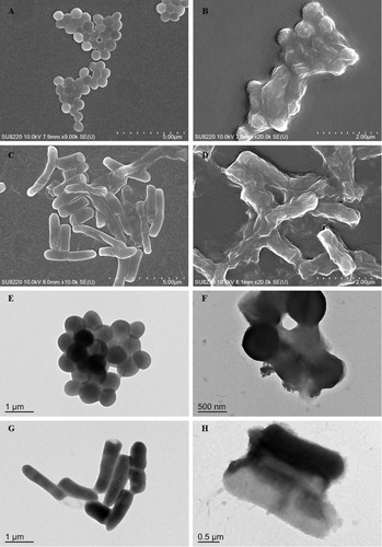
Figure 15 Morphology of the PCC liposomes-treated S. aureus and E. coli observed by SEM and TEM.
Notes: The untreated S. aureus (A, E) and E. coli (C, G), the PCC liposomes-treated S. aureus (B, F) and E. coli (D, H).
Abbreviations: PCC, photo-responsive Camellia sapogenin derivative cationic; SEM, scanning electron microscope; TEM, transmission electron microscope.