Figures & data
Figure 1 (A) TEM image, (B) size distribution graph, (C, D) HRTEM images, (E) XRD pattern, and (F) hysteresis loop of synthesized Mn-IONPs.
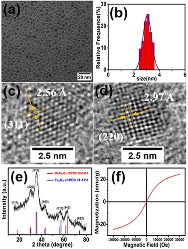
Figure 2 In vitro MR images of the Mn-IONPs-TMAH and Mn-IONPs@PEG (1:1, 1:5, 1:20, and 1:40) at 3.0 T: (A) T1-weighted, (B) T1 mapping, (C) T2-weighted and (D) T2 mapping. (E, F) Linear fitting of 1/T1 and 1/T2 over different (Fe+Mn) concentrations of the nanocomposites.
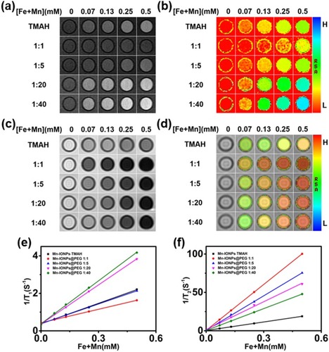
Figure 3 Viability of HepG2 cells after incubation with Mn-IONPs@PEG (1:20) at different concentrations (1–50 µg/mL of [Fe+ Mn]) at 37 °C for 12 hrs and 24 hrs.
![Figure 3 Viability of HepG2 cells after incubation with Mn-IONPs@PEG (1:20) at different concentrations (1–50 µg/mL of [Fe+ Mn]) at 37 °C for 12 hrs and 24 hrs.](/cms/asset/dc20e7ae-8bd6-4a41-9887-61bc7b1b6f08/dijn_a_219749_f0003_b.jpg)
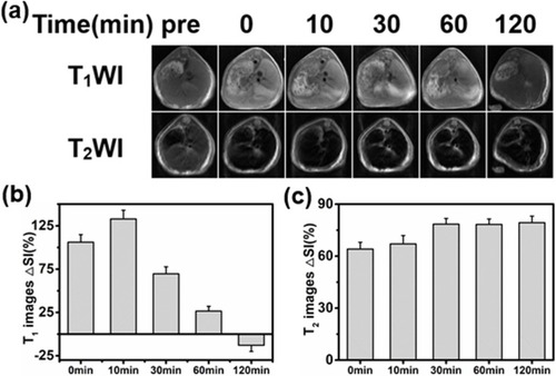
Figure 4 (A) T1-weighted and T2-weighted MR images of transverse planes of the liver using the FSE sequence acquired at 0, 10, 30, 60, or 120 min after intravenous administration of Mn-IONPs@PEG (1:20). (B, C) Enhancement ratio of the MR signal ΔSI of the liver after contrast-enhancement by Mn-IONPs@PEG (1:20).