Figures & data
Figure 1 Most commonly used nanomaterials in nanomedicine manufactured from different substances.
Note: The figure was produced by smart servier medical art library in combination with ChemDrew.
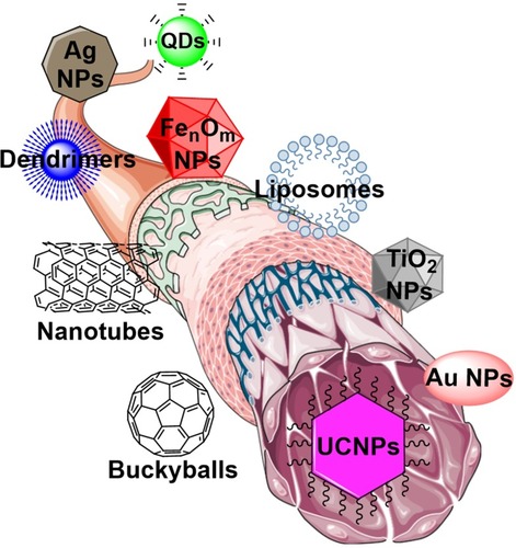
Figure 2 Schematic presentation of UCNPs-PEG-based prodrug with targeting agent.
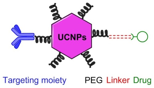
Figure 3 General energy states diagram of the lanthanide ions doped in a low-symmetry crystal.Citation52,Citation53
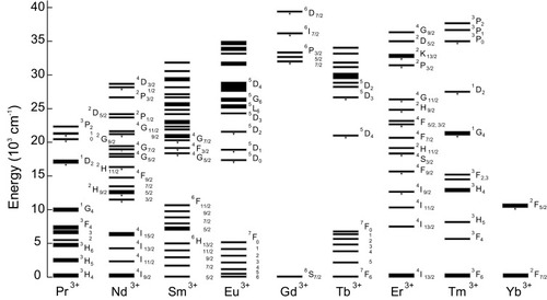
Figure 4 (A) Schematic illustrations of the typical structures of epitaxial and non-epitaxial shells for core-shell tuned UCNPs. Reproduced with permission from Chen X, Peng D, Ju Q, Wang F. Photon upconversion in core-shell nanoparticles. Chem Soc Rev. 2015;44(6):1318–1330.Citation88 Copyright the Royal Society of Chemistry 2015. (B) Schematic illustration of synthesizing and coating process for multilayer NaYF4:Yb,Tm nanoparticles and TEM images from each step. Reproduced with permission from Su Q, Han S, Xie X, et al. The effect of surface coating on energy migration-mediated upconversion. J Am Chem Soc. 2012;134 (51):20849–20857.Citation96 Copyright American Chemistry Society 2012. (C) The left panel shows high-angle annular dark-field micrograph of a NaYF4:Yb,Er/NaGdF4 nanoparticle with the chemical maps from the presence of yttrium (Y) in the core and gadolinium (Gd) in the shell of nanocrystal. The right panel shows the electron energy loss spectroscopy of Y and Gd edges from the nanoparticle with 2.4 nm shell. Reproduced with permission from Zhang F, Che R, Li X, et al. Direct imaging the upconversion nanocrystal core/shell structure at the subnanometer level: shell thickness dependence in upconverting optical properties. Nano Lett. 2012;12(6):2852–2858.Citation98 Copyright American Chemistry Society 2012.
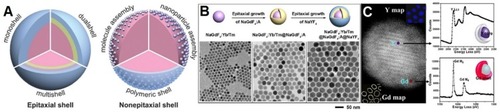
Figure 5 (A) Schematic procedure of synthesizing α-NaYF4:Yb,Er nanocrystals and growing stages consecutively through the thermal decomposition method. Reproduced with permission from Mai H-X, Zhang Y-W, Sun L-D, Yan C-H. Size- and phase-con-trolled synthesis of monodisperse NaYF4: Yb,ErNanocrystals from a unique delayed nucleation pathway monitored with upconversion spectroscopy. J Phys Chem C. 2007;111(37):13730–13739.Citation106 Copyright American Chemical Society 2007. Various sizes of NaYF4-based nanoparticles that have been synthesized under 330°C in OA/ODE (1/1) for (B) 15 min, (C) 25 min, and (D) 45 min. Reproduced with permission from Mai H-X, Zhang Y-W, Si R, et al. High-quality sodium rare-earth fluoride nanocrystals: controlled synthesis and optical properties. J Am Chem Soc. 2006;128(19):6426–6436.Citation107 Copyright American Chemical Society 2006.
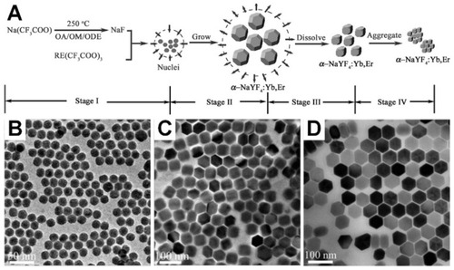
Table 1 Experimental cell viability results of UCNPs under different conditions
Figure 6 (A) Luminescence spectrum of the UCNPs excited at 980 nm and TEM image of the UCNPs. (B) Cell imaging using UCNPs (top left) brightfield image of a cell with internalized UCNPs, (top middle) fluorescence imaging of stained cell with UCNPs excited at 980 nm, and (top right) merged. (Bottom left) brightfield image of a cell without UCNPs, (bottom middle) fluorescence imaging of cell without UCNPs, and (bottom right) cellular autofluorescence excited at 532 nm. (A, B) Reproduced from Wu S, Han G, Milliron DJ, et al. Non-blinking and photostable upconverted luminescence from single lanthanide-doped nanocrystals. Proc Natl Acad Sci. 2009;106(27):10917–10921.Citation153 (C) The fluorescent microscopy images from the trapped droplets by microfluidic array involving: iI) droplets with lanthanide-doped NPs, ii) droplets with GFP-expressing cell, iii) droplets with co-encapsulated NPs and GFP-expressing cell, and iv) empty droplets. Reprinted by permission from Springer Nature: Anal Bioanal Chem, Luminescent nanomaterials for droplet tracking in a microflui- dic trapping array, Vaithiyanathan M, R Bajgiran K, Darapaneni P, Safa N, Dorman JA, Melvin AT, Copyright 2019, doi:10.1007/s00216-018-1448-1.Citation154
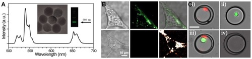
Figure 7 The top scheme indicates fabrication of UCNPs@CCPEB and loading the siRNA process. The bottom scheme indicates the procedure of releasing the siRNA and PDT simultaneously under NIR excitation and UV/visible emission from UCNPs. Reproduced with permission from Zhao H, Hu W, Ma H, et al. Photo-induced charge-variable conjugated polyelectrolyte brushes encapsulating upconversion nanoparticles for promoted siRNA release and collaborative photodynamic therapy under NIR light irradiation. Reproduced from Zhao H, Hu W, Ma H, et al. Photo-induced charge-variable conjugated polyelectrolyte brushes encapsulating upconversion nanoparticles for promoted siRNA release and collaborative photodynamic therapy under NIR light irradiation. Adv Funct Mater. 2017;27(44):1702592Citation170 Copyright John Wiley and Sons.
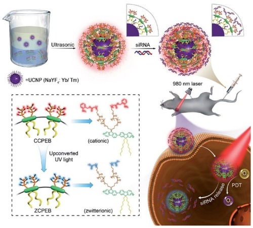
Figure 8 (A) Surface modified UCNPs with organic dyes doped within silicon to get single emission from NPs. Antibody conjugation of these modified nanoparticles creates influential probes for multiplexed in situ molecular mapping of tumor biomarkers. Reproduced with permission from Zhou L, Wang R, Yao C, et al. Single-band upconversion nanoprobes for multiplexed simultaneous in situ molecular mapping of cancer biomarkers. Nat Commun. 2015;6:6938.Citation172 Copyright Springer Nature 2015. (B) Schematic representation of prodrug activation using NIR and intracellular apoptosis by UCL. Reproduced with permission from Min Y, Li J, Liu F, Yeow EKL, Xing B. Near-infrared lightmediated photoactivation of a platinum antitumor prodrug and simultaneous cellular apoptosis imaging by upconversion-luminescent nanoparticles. Angew Chem Int Ed. 2014;53(4):1012–1016.Citation173 Copyright 2013, John Wiley and Sons.
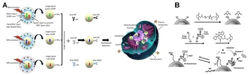
Figure 9 (A) Schematic illustration of microenvironment-sensitive strategy. Triggering the accumulation of UCNPs into the tumor. (B) Mice treated with PDT agent under NIR (G 1), control agent under NIR (G 2), PDT agent without NIR (G 3), and saline under NIR (G 4). Reproduced with permission from Ai X, Ho CJH, Aw J, et al. In vivo covalent cross-linking of photon-converted rare-earth nanostructures for tumour localization and theranostics. Nat Commun. 2016;7:10432.Citation184 (C) In vivo UCL imaging of a tumor-bearing mouse after intratumoral injection of UCNPs solution into the tumor site. Reproduced with permission from Dai Y, Xiao H, Liu J, et al. In vivo multimodality imaging and cancer therapy by near-infrared light-triggered trans-platinum pro- drug-conjugated upconverison nanoparticles. J Am Chem Soc. 2013;135(50):18920–18929.Citation185 Copyright American Chemistry Society 2013.
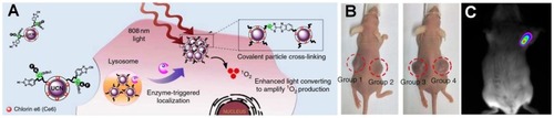
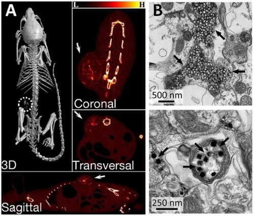
Figure 10 (A) X-ray CT images of the tumor-bearing mice after intratumoral injection of NP/PDA suspension. Reproduced with permission from Dai Y, Yang D, Yu D, et al. Mussel-inspired polydopamine-coated lanthanide nanoparticles for NIR-II/CT dual imaging and photothermal therapy. ACS Appl Mater Interfaces. 2017;9(32):26674– 26683.Citation186 Copyright American chemical society 2017. (B) Electron micrographs of UCNPs distributed in the neuronal tissue. Black arrows indicate clusters of UCNPs. Top image shows the distribution of most UCNPs in extracellular space, and the bottom image shows the uptake of UCNPs within an axon. From Chen S, Weitemier AZ, Zeng X, et al. Near-infrared deep brain stimulation via upconversion nanoparticle–mediated optogenetics. Science. 2018;359(6376):679.Citation187 Reprinted with permission from AAAS https://science.sciencemag.org/content/359/6376/679.