Figures & data
Figure 1 Amperograms (current calibrated as concentration vs time) of NO and ONOO− release stimulated by LDL with different patterns on the surface of endothelial cells. a) NO release from endothelial cells stimulated by LDL (Patterns A, B, and I, 1,000 µg/mL). b) ONOO− release from endothelial cells stimulated by LDL (Patterns A, B, and I, 1,000 µg/mL). Arrows indicate LDL injection.
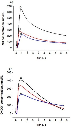
Figure 2 Maximal NO and ONOO− release from the surface of endothelial cells stimulated by LDL with different patterns. a) Maximal NO and ONOO− release from endothelial cells stimulated by LDL (Patterns A, B, and I, 1,000 µg/mL), solid bar indicates NO and open bar indicates ONOO−. b) A ratio of maximal NO to ONOO−. Data are expressed as mean±SD. Significance was determined using Student’s t-test. *P<0.01 vs B.
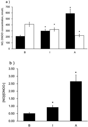
Figure 3 Dose-dependent NO and ONOO− release from the surface of endothelial cells stimulated by LDL. a) Production of NO stimulated by LDL with different patterns (A, B, and I) and different concentrations (from 50 µg/mL to 1,000 µg/mL). b) Production of ONOO− stimulated by LDL with different patterns (A, B, and I) and different concentrations (from 50 µg/mL to 1,000 µg/mL). c) The ratio of NO to ONOO−. Black triangle, white circle, and black dot indicate LDL injection of pattern A, B, and I, respectively.
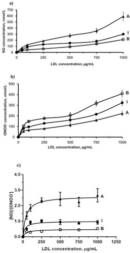
Figure 4 NO and ONOO− release stimulated by LDL mixture with different combinations. a) NO and ONOO− production stimulated by LDL mixture (800 µg/mL). Solid bar indicates NO and open bar indicates ONOO−. b) Ratio of NO to ONOO−. Data are expressed as mean±SD.
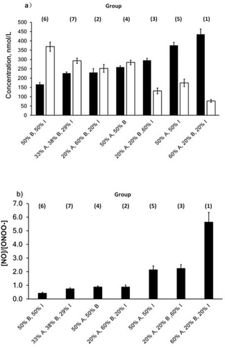
Figure 5 NO and ONOO− release from endothelial cells stimulated by LDL after incubation with different treatments. Endothelial cells were incubated with control EBM, PEG-SOD (400 U/mL), L-arginine (300 µM), sepiapterin (200 µM), L-NAME (100 µM), and VAS2870 (10 µM) at 37ºC for 30 minutes. a) NO production stimulated by LDL (Patterns A, B, and I, 800 µg/mL). b) ONOO− production stimulated by LDL (Patterns A, B, and I, 800 µg/mL). c) Ratio of NO to ONOO−. Data are expressed as mean±SD.
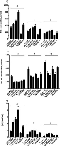
Figure 6 NO and ONOO− release stimulated by ox-LDL/n-LDL. a) NO production stimulated by ox-LDL/n-LDL (800 µg/mL). b) ONOO− production stimulated by ox-LDL/n-LDL (800 µg/mL). c) Ratio of NO to ONOO−. Data are expressed as mean±SD. Significance was determined using Student’s t-test. *P<0.01 vs n-LDL.
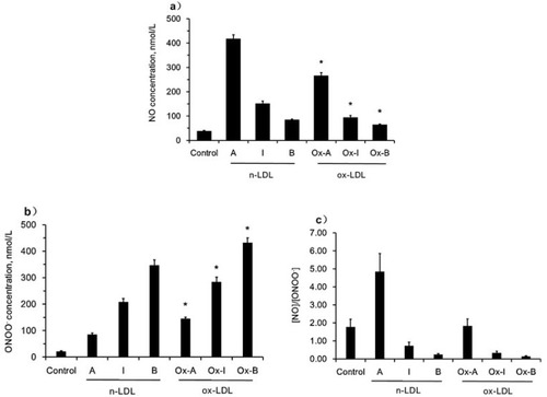
Figure 7 Monocyte adhesion and cell adhesion molecule expression stimulated by n-LDL. a) Monocyte adhesion stimulated by subclass A, B, and I of n-LDL (400 µg/mL) measured at different incubation time (from 10 to 60 minutes). b) Dose-dependent monocyte adhesion stimulated by patterns A, B, and I of n-LDL (50, 100, 200, 400 µg/mL). Data are expressed as mean±SD. MFI indicates mean fluorescence intensity. c) Effect of LDL with different patterns on the expression of ICAM-1 and VCAM-1. Endothelial cells were incubated with LDL of patterns A, B, and I (400 µg/mL) at 37ºC for 5 hours. After incubation, the cells were washed with DPBS and fixed with 4% formaldehyde solution. ICAM-1 and VCAM-1 expression were determined by cell ELISA. Data are expressed as mean±SD. Solid bar indicates ICAM-1, open bar indicates VCAM-1. OD indicates optical density. Significance was determined using Student’s t-test. *P<0.01 vs control.
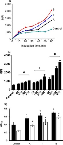
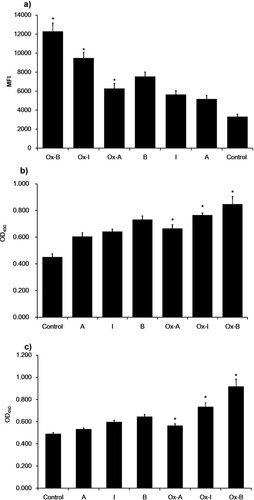
Figure 8 Monocyte adhesion and cell adhesion molecular expression stimulated by n-LDL or ox-LDL. a) Monocyte adhesion stimulated by n-LDL or ox-LDL of patterns A, B, and I (400 µg/mL). Data are expressed as mean±SD. MFI indicates mean fluorescence intensity. b) Effect of n-LDL/ox-LDL with different patterns on the expression of ICAM-1. Endothelial cells were incubated with n-LDL/ox-LDL of patterns A, B, and I (400 µg/mL) at 37ºC for 5 hours. After incubation, the cells were washed with DPBS and fixed with 4% formaldehyde solution. ICAM-1 and VCAM-1 expression were determined by cell ELISA. c) Effect of n-LDL or ox-LDL with different subclasses on the expression of VCAM-1. Data are expressed as mean±SD. OD450 indicates optical density. Significance was determined using Student’s t-test. *P<0.01 vs n-LDL.