Figures & data
Figure 1 Overall synthesis of monodisperse PEG/Ca(II)/MNGs.
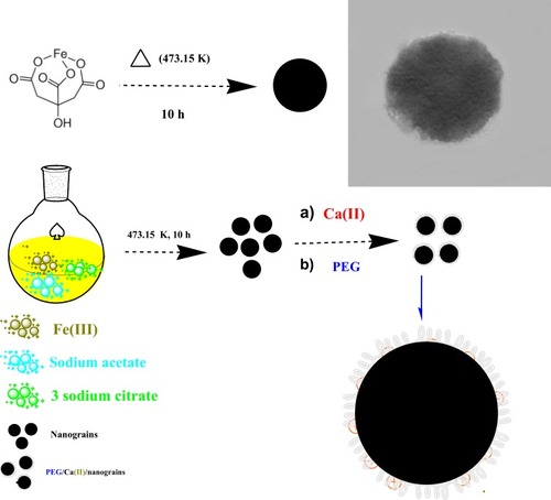
Figure 2 (A) FT-IR spectra of MNGs and PEG/Ca(II)/MNGs. (B) Zeta potential of MNGs and PEG/Ca(II)/MNGs. (C) Hysteresis loops of MNGs and PEG/Ca(II)/MNGs. In the inset: photograph of an aqueous PEG/Ca(II)/MNGs in a vial without/with magnetic field. (D) XRD pattern of bare MNGs. In the inset: SAED pattern of MNGs.
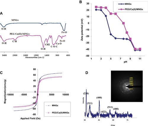
Figure 3 (A) SEM of bare MNGs. (B) TEM of bare MNGs; inset: size distribution of MNGs.
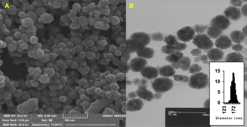
Figure 4 (A) Size distribution of bare MNGs and PEG/Ca(II)/MNGs. (B) TEM image of PEG/Ca(II)/MNGs. (C) SEM image of PEG/Ca(II)/MNGs (scale bar is 50 nm).
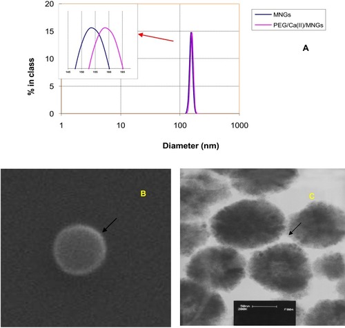
Figure 5 (A) Viability assay of E. coli in three incubation times. (B) Relation between nanocarrier concentration and number of colonies. (C) Relation between nanocarrier concentration variations in different gene delivery steps and colony number. (D) Effect of LB volume on the colony number. The colony number evaluation of transformed bacteria cells, (E) control, (F) group 2, (G) group 1. The blue colonies show viable and transformed bacteria.
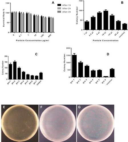
Figure 6 (A) Agarose gel electrophoresis of extracted plasmid. Lane 1: molecular marker, Lane 2: treated, Lane 3: control: (B) Restriction endonuclease digestion. Lane 1: molecular marker, Lane 2: enzyme digested pDNA extracted from treated-cells, Lane 3: enzyme digested pDNA extracted from control cells. (C) Agarose gel electrophoresis of NeoR gene (807 bp amplicon). Lane 1: DNA molecular weight marker; Lane 2: treated sample; Lane 3: control.
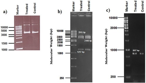
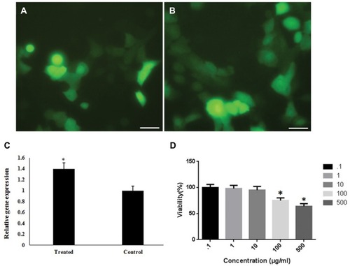
Figure 7 Gene delivery efficiency of GFP by PEG/Ca(II)/MNGs: (A) Phase contrast fluorescence microscopy image of transfected cells in the Control group. (B) Fluorescence image of transfected GFP in cells by PEG/Ca(II)/MNGs. (C) Relative GFP expression by qPCR in treated cell by PEG/Ca(II)/MNGs and lipofectamine 2000 as a standard method (Scale bar is 20 µm). (D) Viability of exposed HEK 293T cells to PEG/Ca(II)/MNGs. All experiments were performed in triplicate.